| Correction to: Desk of the Editor Vol. 9 Issue 4 Due to an unfortunate error in the email address of the corresponding author, this correction paper is being published. |
Donut Mastopexy LumpectomyAbstractAdvancements in oncoplastic techniques have enhanced commitment to restore shape and, hence, has improved cosmetic outcomes. Donut mastopexy lumpectomy is one such technique and is best utilized in a setting of a malignancy not extending to the skin or the nipple-areolar complex. As a potential alternative to standard lumpectomy, it has many advantages including restriction of scar to the periareolar region, ease and rapidity of surgery, retention of nipple-areolar sensation, and the possibility of performing augmentation mammoplasty. A mini breast lift is also provided without ugly and visible scars. This report provides an insight into the technical details and utility of donut mastopexy lumpectomy (DML) in breast oncoplasty. |
| Correction to: Assessment Awareness of Public About Breast Cancer and its Screening Measurements in Asir Region, KSA With the author(s)' decision to step back from Open Choice, the copyright of the article changed on April 2019 to © Indian Association of Surgical Oncology 2019 and the article is forthwith distributed under the terms of copyright. |
Isolated Midbrain Metastasis from Breast Cancer: a Classic Spotter DiagnosisAbstractIsolated midbrain metastasis from breast cancer is a rare occurrence. We present a classical clinical image of a woman who presented with breast cancer with synchronous isolated midbrain metastasis. |
A Leiomyosarcoma of Inferior Vena Cava Presenting as a Liver Metastasis Mass in a Patient with History of Transitional Cell CarcinomaAbstractThe most probable diagnosis for a newly detected mass in the cancer patients is secondary metastasis. However, the multiple primary tumors should not be off the table of diagnoses. In this study, a 70-year-old man with the history of transitional cell carcinoma (TCC) was reported who had been referred due to a newly detected mass in the hepatic segment one which adhered to the inferior vena cava (IVC). Although the most probable diagnosis according to the patient's medical history was secondary metastasis, the biopsy revealed a leiomyosarcoma (LMS) tumor. Therefore, a mass biopsy can be determinative for confirming the diagnosis and further management of cancer patients with a newly detected mass. |
Primary Malignant Peripheral Nerve Sheath Tumor of the Trachea: a Case Report with Brief Review of LiteratureAbstractMalignant schwannoma, also called malignant peripheral nerve sheath tumor (MPNST), is a rare and aggressive tumor arising from the nerve sheath. We describe a rare case of endotracheal malignant peripheral nerve sheath tumor occurring in a middle-aged male who presented with asthma-like symptoms for 6 months with progressively increasing dyspnea. A computed tomogram (CT) scan of the thorax revealed near complete luminal obstruction of the trachea by a mass lesion at the level of the second and third tracheal rings. Microlaryngotracheoscopy revealed a fleshy pedunculated growth arising from the left side of the second and third tracheal rings and obliterating almost the entire tracheal lumen. Intraluminal complete excision of the mass was done. Later, he underwent excision of the 2nd and 3rd rings after the histopathology revealed MPNST. Patient after 28 months of follow-up is free of disease. |
| Late Presentation of Chronic-Organised Biloma Masquerading as Gallbladder Fossa Mass Years After Cholecystectomy: a Diagnostic Enigma |
Papillary Carcinoma in Thyroglossal Cyst: an Unusual CaseAbstractMalignancy developing in thyroglossal cysts is very rare. Papillary carcinoma is the most common histopathological type of carcinoma encountered in thyroglossal cysts followed by squamous cell carcinoma. A 50-year-old male presented with a midline neck swelling. After ultrasonography and fine needle aspiration cytology, he underwent Sistrunk operation for removal of the thyroglossal cyst. The histopathology was reported as papillary carcinoma of the thyroid. So, he underwent total thyroidectomy, which showed foci of papillary microcarcinoma. Clinical awareness of this rare condition is essential for proper management. Possibility of malignancy arising in thyroglossal cysts should be considered in elderly patients. |
Laparoscopic Ovarian Transposition in Rectal Cancer: More than Just Oncological OutcomesAbstractLocally advanced rectal cancer (LARC) is treated with neoadjuvant chemoradiotherapy which down stages tumor and improves complete resection rates thus reducing local recurrences. Pelvic radiotherapy improves oncological outcomes; however, it is associated with ovarian irradiation and premature menopause. This has a consequence to fertility and hormone preservation in young women diagnosed with locally advanced rectal cancer. Laparoscopic ovarian transposition is an established method to preserve ovarian function. This review discusses the technique, indications, and limitations of laparoscopic ovarian transposition in young women requiring pelvic radiotherapy. |
Swallowing Skills and Aspiration Risk Following Treatment of Head and Neck CancersAbstractSurgical resection and chemoradiation are common modalities of treatment in head and neck cancers. Dysphagia is one of the common complications following these interventions. The severity of dysphagia depends on various factors, site and extent of resection, and radiation therapy to highlight a few. Thirty-five head and neck cancer patients treated with surgical and/or chemoradiation were assessed for parameters of swallowing. Extent of resection was statistically associated with swallowing symptoms phase wise. The results revealed a strong association between the presence of aspiration with resection of the tongue base and radiation therapy (p < 0.01). Oral preparatory and oral phase abnormalities were present in all the cases with varying severity especially in cases where the mandible and body of tongue were compromised (p < 0.05). These findings provide a specific profile which has high clinical utility. |
Saturday, May 25, 2019
Surgical Oncology

Current Treatment Options in Oncology
QT Interval Prolongation Associated With Cytotoxic and Targeted Cancer TherapeuticsOpinion statementCardiovascular toxicities are potentially serious treatment limiting complications of many different cancer therapeutics including traditional cytotoxic chemotherapies as well as targeted- and immunotherapies. As a result, there is increased monitoring for cancer treatment-related cardiotoxicities, ranging from heart failure to arrhythmias. Many anticancer treatments are known to prolong the QT interval through a variety of mechanisms including direct effects on ion channels and indirectly via intracellular signaling pathways. While QT prolongation increases the risk for the potentially life-threatening ventricular arrhythmia torsades de pointes, the incidence of this arrhythmia in the setting of most cancer treatments is quite rare, and the majority of patients can continue safely receiving these medications despite their QT prolonging potential. A multidisciplinary approach to the cardiovascular care of the cancer patient is essential to mitigate risk of cardiotoxicity while minimizing unnecessary treatment disruption of potentially life-saving cancer treatments. |
Pediatric Cardio-Oncology: Development of Cancer Treatment-Related Cardiotoxicity and the Therapeutic Approach to Affected PatientsOpinion statementThe past 5 decades have seen significant improvements in outcomes for pediatric patients with cancer. Unfortunately, children and adolescents who have been treated for cancer are five to six times more likely to develop cardiovascular disease as a result of their therapies. Cardiovascular disease may manifest in a plethora of ways, from asymptomatic ventricular dysfunction to end-stage heart failure, hypertension, arrhythmia, valvular disease, early coronary artery disease, or peripheral vascular disease. A number of treatment modalities are implicated in pediatric and adult populations, including anthracyclines, radiation therapy, alkylating agents, targeted cancer therapies (small molecules and antibody therapies), antimetabolites, antimicrotubule agents, immunotherapy, interleukins, and chimeric antigen receptor T cells. For some therapies, such as anthracyclines, the mechanism of injury is elucidated, but for many others it is not. While a few protective strategies exist, in many cases, observation and close monitoring is the only defense against developing end-stage cardiovascular disease. Because of the variety of potential outcomes after cancer therapy, a one-size-fits-all approach is not appropriate. Rather, a good working relationship between oncology and cardiology to assess the risks and benefits of various therapies and planning for appropriate surveillance is the best model. When disease is identified, any of a number of therapies may be appropriate; however, in the pediatric and adolescent population supportive data are limited. |
Diagnosis and Management of Subcutaneous Soft Tissue SarcomaOpinion statementThe proper diagnosis and treatment planning for subcutaneous soft tissue sarcoma is very important. Soft tissue tumors can occur anywhere in the body, but if they occur subcutaneously, patients can easily notice a subcutaneous soft tissue mass. Therefore, it is possible to determine through recording, the growth speed of the mass, which is often difficult to obtain with deep-situated soft tissue masses. Palpation can also provide information about the firmness and mobility of the mass. Thus, history taking and physical examinations are informative for subcutaneous soft tissue tumors, compared to tumors that occur deeply. Because subcutaneous soft tissue tumors are easily recognized, they are often resected, without sufficient imaging analyses or thorough treatment planning. An operation performed based on such an inadequate preoperative plan is called a "whoops surgery." In the case of "whoops surgeries," subsequent radical surgery is required to remove additional areas, including hematomas that result from the initial surgery, that require a wider range of resection and soft tissue reconstruction. Therefore, as with deep-seated soft tissue tumors, it is important to conduct careful imaging examinations and make appropriate preoperative plans for subcutaneous soft tissue tumors. Subcutaneous soft tissue sarcomas often show an invasive pattern, and such tumors require a more careful assessment to prevent local recurrence after surgery. During surgery, it is necessary to remove the entire infiltration area along the fascia. Sometimes, an adequately wide excision is necessary, which is considered the minimum necessary procedure to eradicate the lesion. As noted above, clinicians who see patients with subcutaneous soft tissue tumors are encouraged to have sufficient knowledge and experience regarding the diagnosis and treatment. This article is intended for all doctors who deal with subcutaneous soft tissue tumors and focuses on essential points regarding their diagnosis and management. |
| Correction to: Immunotherapy Advances in Urothelial Carcinoma In the original version of this article, which published in Current Treatment Options in Oncology, Volume 20, Issue 12, December 2018, the surname of the third author was captured incorrectly. The name shown above is correct. |
The Role of CDK4/6 Inhibitors in Breast CancerOpinion statementOral inhibitors of CDK4/6 have been shown to increase response rates and prolong disease control when combined with endocrine therapy in hormone-responsive (HR+) HER2-negative advanced breast cancer. Palbociclib, ribociclib and abemaciclib are all approved in combination with non-steroidal aromatase inhibitors in first-line therapy for post-menopausal women, with a 40–45% improvement in progression-free survival seen with the addition of any of these CDK4/6 inhibitors. Additional approved indications, including first- and second-line combination therapy for pre-menopausal women, combination with fulvestrant and use as monotherapy, vary with each agent and are reviewed fully in the subsequent texts. These agents also differ in their toxicity profiles and monitoring requirements, and prescribers should be aware of the individual requirements for each agent. Current clinical trials are investigating the expanded use of these agents in other breast cancer subtypes, such as HER2-positive and triple-negative breast cancer, as well as in the adjuvant and neoadjuvant treatments of early breast cancer. Resistance to CDK4/6 inhibition can occur through multiple mechanisms. Rational combinations with other therapies, such as PI3K inhibitors, HER2-directed therapies and immunotherapy, are being explored. |
Multimodality Therapy of Patients with Refractory MeningiomasOpinion statementRecurrent and refractory meningiomas are a clinical challenge and treatment at the time of recurrence is not well delineated. Treatment with surgery and/or radiation remain the mainstay, but each has their limitations and risks. The search for an adjuvant systemic therapy continues and as many of the initially promising approaches have not had reproducible responses. Bevacizumab has shown some efficacy in controlling recurrent disease and could be useful in disease that is multifocal or in close proximity to critical structures. Other targeted therapies, as well as immunotherapy, are being studied and trials are in development. Though we are hopeful that these novel therapies will benefit patients with refractory meningiomas, we approach them with some trepidation. This is due to prior failures of immunotherapy and targeted therapy in central nervous system disease. In addition, there is known difficulty in developing trials and assessing response with these slow-growing tumors. |
Cardiotoxicity of Contemporary Breast Cancer TreatmentsOpinion statementTreatment-related cardiotoxicity remains a significant concern for breast cancer patients undergoing cancer treatment and extends into the survivorship period, with adverse cardiovascular (CV) outcomes further compounded by the presence of pre-existing CV disease or traditional CV risk factors. Awareness of the cardiotoxicity profiles of contemporary breast cancer treatments and optimization of CV risk factors are crucial in mitigating cardiotoxicity risk. Assessment of patient- and treatment-specific risk with appropriate CV surveillance is another key component of care. Mismatch between baseline cardiotoxicity risk and intensity of cardiotoxicity surveillance can lead to unnecessary downstream testing, increased healthcare expenditure, and interruption or discontinuation of potentially life-saving treatment. Efforts to identify early imaging and/or circulating biomarkers of cardiotoxicity and develop effective management strategies are needed to optimize the CV and cancer outcomes of breast cancer survivors. |
Cancer and Coronary Artery Disease: Common Associations, Diagnosis and Management ChallengesOpinion statementCoronary artery disease (CAD) and cancer often occur in the same patients via common biological pathways and shared risk factors. A variety of chemotherapeutic agents and radiotherapy can influence the development and progression of CAD. The diagnosis of ischaemic heart disease may be challenging in certain cases such as premature CAD secondary to radiotherapy. The management of CAD in cancer patients in the stable, acute and chronic settings can often be complicated by issues related to ongoing or previous cancer treatment or the cancer itself. A multidisciplinary approach in the setting of a cardio-oncology service is often best-served to optimally treat such patients. |
Pain in Cancer Survivors: How to ManageOpinion statementManaging pain in cancer survivors requires that oncologists understand the common painful syndromes that can occur from treatment or disease. Assessment no longer singularly focuses on pain characteristics (e.g., intensity, quality, location), now incorporating a strong focus on functional impairment and potential improvement that might occur with adequate treatment. Improvement in function is now the goal used to measure success. In addition, assessment must incorporate risk factors that might predispose patients to substance use disorder so that interventions can be implemented to mitigate this risk. Universal precautions are measures that help assess and ensure adherence to the treatment plan and may include the use of agreements, urine toxicology, and review of dispensing information derived from state prescription drug monitoring program (PDMP). These are generally obtained annually for all individuals, although some states have instituted mandatory review of the PDMP whenever prescribing an opioid. For patients at moderate to high risk for misuse of opioids, where opioids are warranted for the treatment of their pain syndrome, universal precautions are instituted more frequently. Other measures may include prescribing a 1- to 2-week supply of medications if compulsive use leads the patient to running out of drug early, and in some cases, family members may be employed to dispense daily allotments of the medication. When opioids are no longer indicated, gradual tapering of the drug by approximately 10% per month is generally sufficient to prevent withdrawal symptoms and ensure patient acceptance. |
Malignant Melanoma: Autoimmunity and Supracellular Messaging as New Therapeutic ApproachesOpinion statementMelanoma is one of the most aggressive forms of cancer, with a high mortality rate in the absence of a safe and curable therapy. As a consequence, several procedures have been tested over time, with the most recent (immunological and targeted) therapies proving to be effective in some patients. Unfortunately, these new treatment options continue to generate debate related to the therapeutic strategy (intended to maximize the long-term results of patients with melanoma), not only about the monotherapy configuration but also regarding association/succession between distinct therapeutic procedures. As an example, targeted therapy with BRAF inhibitors proved to be effective in advanced BRAF-mutant melanoma. However, such treatments with BRAF inhibitors lead to therapy resistance in half of patients after approximately 6 months. Even if most benign nevi incorporate oncogenic BRAF mutations, they rarely become melanoma; therefore, targeted therapy with BRAF inhibitors should be viewed as an incomplete or perfectible therapy. Another example is related to the administration of immune checkpoint inhibitors/ICIs (anti-CTLA-4 antibodies, anti-PD-1/PD-L1 antibodies), which are successfully used in metastatic melanoma. It is currently believed that CTLA-4 and PD-1 blockade would favor a strong immune response against cancer cells. The main side effects of ICIs are represented by the development of immune-related adverse events, which in some cases can be lethal. These ICI side effects would thus be not only therapeutically counterproductive but also potentially dangerous. Surprisingly, a subset of immune-related adverse events (especially autoimmune toxicity) seems to be clearly correlated with better therapeutic results, perhaps due to an additional therapeutic effect (currently insufficiently studied/exploited). Contrary to the classical approach of cancer (considered until now an uncontrolled division of cells), a very recent and comprehensive theory describes malignancy as a supracellular disease. Cancerous disease would therefore be a disturbed supracellular process (embryogenesis, growth, development, regeneration, etc.), which imposes/coordinates an increased rhythm of cell division, angiogenesis, immunosuppression, etc. Melanoma is presented from such a supracellular perspective to be able to explain the beneficial role of autoimmunity in cancer (autoimmune abortion/rejection of the melanoma-embryo phenotype) and to create premises to better optimize the newly emerging therapeutic options. Finally, it is suggested that the supracellular evolution of malignancy implies complex supracellular messaging (between the cells and host organism), which would be interfaced especially by the extracellular matrix and noncoding RNA. Therefore, understanding and manipulating supracellular messaging in cancer could open new treatment perspectives in the form of digitized (supracellular) therapy. |
Environmental Sciences
| Effect of amendments on the leaching behavior of alkaline anions and metal ions in bauxite residue Publication date: November 2019 Source: Journal of Environmental Sciences, Volume 85 Author(s): Tao Tian, Jingju Zhou, Feng Zhu, Yuzhen Ye, Ying Guo, William Hartley, Shengguo Xue AbstractA column leaching experiment was used to investigate the efficacy of amendments on their ability to remove alkaline anions and metal ions from bauxite residue leachates. Treatments included, simulated acid rain (AR), phosphogypsum + vermicompost (PVC), phosphogypsum + vermicompost + simulated acid rain (PVA), and biosolids + microorganisms (BSM) together with controls (CK). Results indicated that amendment could effectively reduce the leachate pH and EC values, neutralize OH−, CO32−, HCO3−, and water soluble alkali, and suppress arsenic (As) content. Correlation analysis revealed significant linear correlations with pH and concentrations of OH−, CO32−, HCO3−, water-soluble alkali, and metal ions. BSM treatment showed optimum results with neutralizing anions (OH−, CO32−, and HCO3−), water soluble alkali, and removal of metal ions (Al, As, B, Mo, V, and Na), which was attributed to neutralization from the generation of small molecular organic acids and organic matter during microbial metabolism. BSM treatment reduced alkaline anions and metal ions based on neutralization reactions in bauxite residue leachate, which reduced the potential pollution effects from leachates on the soil surrounding bauxite residue disposal areas. Graphical abstract |
| Arsenic mobilization from soils in the presence of herbicides Publication date: November 2019 Source: Journal of Environmental Sciences, Volume 85 Author(s): Yuxuan Jiang, Wen Zhong, Wei Yan, Li Yan AbstractArsenic (As) mobilization in soils is a fundamental step controlling its transport and fate, especially in the presence of the co-existing components. In this study, the effect of two commonly used herbicides, glyphosate (PMG) and dicamba, and two competing ions including phosphate and humic acid, on As desorption and release was investigated using batch and column experiments. The batch kinetics results showed that As desorption in the presence of competing factors conformed to the pseudo-second order kinetics at pH range of 5–9. The impact of phosphate on desorption was greatest, followed by PMG. The competitive effect of dicamba and humic acid was at the same level with electrolyte solution. In situ flow cell ATR-FTIR analysis was performed to explore the mechanism of phosphate and PMG impact on As mobilization. The results showed that PMG promoted As(III) desorption by competiting for available adsorption sites with no change in As(III) complexing structure. On the other hand, phophate changed As(III) surface complexes from bidentate to monodentate structures, exhibiting the most siginficant effect on As(III) desorption. As(V) surface complexes remained unchanged in the presence of PMG and phosphate, implying that the competitive effect for As(V) desorption was primarily determined by the available adsorption sites. Long-term (10 days) soil column experiments suggested that the effect of humic acid on As mobilization became pronounced from 3 days (18 PVs). The insights of this study help us understand the transport and fate of As due to herbicides application. Graphical abstract |
| Bacterial resistance to lead: Chemical basis and environmental relevance Publication date: November 2019 Source: Journal of Environmental Sciences, Volume 85 Author(s): Qiying Nong, Ke Yuan, Zhuang Li, Ping Chen, Yongshun Huang, Ligang Hu, Jie Jiang, Tiangang Luan, Baowei Chen AbstractNatural bacterial isolates from heavily contaminated sites may evolve diverse tolerance strategies, including biosorption, efflux mechanism, and intracellular precipitation under the continually increased stress of toxic lead (Pb) from anthropogenic activities. These strategies utilize a large variety of functional groups in biological macromolecules (e.g., exopolysaccharides (EPSs) and metalloproteins) and inorganic ligands, including carboxyl, phosphate and amide groups, for capturing Pb. The amount and type of binding sites carried by biologically originated materials essentially determines their performance and potential for Pb removal and remediation. Many factors, e.g., metal ion radius, electronegativity, the shape of the cell surface sheath, temperature and pH, are thought to exert significant influences on the abovementioned interactions with Pb. Conclusively, understanding the chemical basis of Pb-binding in these bacteria can allow for the development of effective microbial Pb remediation technologies and further elucidation of Pb cycling in the environment. Graphical abstract |
| Effects of imazethapyr spraying on plant growth and leaf surface microbial communities in Arabidopsis thaliana Publication date: November 2019 Source: Journal of Environmental Sciences, Volume 85 Author(s): Wanyue Liu, Mingjing Ke, Zhenyan Zhang, Tao Lu, Youchao Zhu, Yan Li, Xiangliang Pan, Haifeng Qian AbstractImazethapyr (IM) is an acetolactate synthase (ALS)-inhibiting herbicide that has been widely used in recent years. However, IM spraying can lead to the accumulation of herbicide residues in leaves. Here, we determined the effects of IM spraying on the plant growth and leaf surface microbial communities of Arabidopsis thaliana after 7 and 14 days of exposure. The results suggested that IM spraying inhibited plant growth. Fresh weight decreased to 48% and 26% of the control value after 7 and 14 days, respectively, of 0.035 kg/ha IM exposure. In addition, anthocyanin content increased 9.2-fold and 37.2-fold relative to the control content after 7 and 14 days of treatment, respectively. Furthermore, IM spraying destroyed the cell structures of the leaves, as evidenced by increases in the number of starch granules and the stomatal closure rate. Reductions in photosynthetic efficiency and antioxidant enzyme activity were observed after IM spraying, especially after 14 days of exposure. The diversity and evenness of the leaf microbiota were not affected by IM treatment, but the composition of community structure at the genus level was altered by IM spraying. Imazethapyr application increased the abundance of Pseudomonas, a genus that includes species pathogenic to plants and humans, indicating that IM potentially increased the abundance of pathogenic bacteria on leaves. Our findings increase our understanding of the relationships between herbicide application and the microbial community structures on plant leaves, and they provide a new perspective for studying the ecological safety of herbicide usage. Graphical abstract |
| Fate of antibiotics and antibiotic resistance genes in a full-scale restaurant food waste treatment plant: Implications of the roles beyond heavy metals and mobile genetic elements Publication date: November 2019 Source: Journal of Environmental Sciences, Volume 85 Author(s): Pinjing He, Zhuofeng Yu, Liming Shao, Yizhou Zhou, Fan Lü AbstractIs our food safe and free of the crisis of antibiotics and antibiotic resistance (AR)? And will the derived food waste (FW) impose AR risk to the environment after biological treatment? This study used restaurant FW leachates flowing through a 200 tons-waste/day biological treatment plant as a window to investigate the fate of antibiotics and antibiotic-resistance genes (ARGs) during the acceptance and treatment of FW. Sulfonamides (sulfamethazine, sulfamethoxazole) and quinolones (ciprofloxacin, enrofloxacin, ofloxacin) were detected during FW treatment, while tetracyclines, macrolides and chloramphenicols were not observable. ARGs encoding resistance to sulfonamides, tetracyclines and macrolides emerged in FW leachates. Material flow analysis illustrated that the total amount of antibiotics (except sulfamethazine) and ARGs were constant during FW treatment processes. Both the concentration and total amount of most antibiotics and ARGs fluctuated during treatment, physical processes (screening, centrifugation, solid–liquid and oil–water separation) did not decrease antibiotic or ARGs concentrations or total levels permanently; the affiliated wastewater treatment plant appeared to remove sulfonamides and most ARGs concentrations and total amount. Heavy metals Ni, Co and Cu were important for disseminating antibiotics concentrations and MGEs for distributing ARGs concentrations. Humic substances (fulvic acids, hydrophilic fractions), C-associated and N-associated contents were essential for the distribution of the total amounts of antibiotics and ARGs. Overall, this study implied that human food might not be free of antibiotics and ARGs, and FW was an underestimated AR pool with various determinants. Nonetheless, derived hazards of FW could be mitigated through biological treatment with well-planned daily operations. Graphical abstract |
| Effects of typical algae species (Aphanizomenon flosaquae and Microcystis aeruginosa) on photoreduction of Hg2+ in water body Publication date: November 2019 Source: Journal of Environmental Sciences, Volume 85 Author(s): Rongguo Sun, Yafei Mo, Xinbin Feng, Leiming Zhang, Lin Jin, Qiuhua Li AbstractPhotoreduction characteristics of divalent inorganic mercury (Hg2+) in the presence of specific algae species are still not well known. Laboratory experiments were conducted in the present study to identify the effects of different concentrations of living/dead algae species, including Aphanizomenon flosaquae (AF) and Microcystis aeruginosa (MA), on the photoreduction rate of Hg2+ under various light conditions. The experimental results showed that percentage reduction of Hg2+ was significantly influenced by radiation wavelengths, and dramatically decreased with the presence of algae. The highest percentage reduction of Hg2+ was induced by UV-A, followed by UV-B, visible light and dark for both living and dead AF, and the order was dark > UV-A > UV-B > visible light for both living and dead MA. There were two aspects, i.e., energy and attenuation rate of light radiation and excrementitious generated from algae metabolisms, were involved in the processes of Hg2+ photoreduction with the presence of algae under different light conditions. The percentage reduction of Hg2+decreased from 15% to 11% when living and dead AF concentrations increased by 10 times (from 106 to 105 cells/mL), and decreased from 11% to ~ 9% in the case of living and dead MA increased. Algae can adsorb Hg2+and decrease the concentration of free Hg2+, thus inhibiting Hg2+ photoreduction, especially under the conditions with high concentrations of algae. No significant differences were found in percentage reduction of Hg2+between living and dead treatments of algae species. The results are of great importance for understanding the role of algae in Hg2+ photoreduction. Graphical abstract |
| Exposure to low-level metalaxyl impacts the cardiac development and function of zebrafish embryos Publication date: November 2019 Source: Journal of Environmental Sciences, Volume 85 Author(s): Yuqiong Wu, Ying Zhang, Meng Chen, Qihong Yang, Shanshan Zhuang, Liangju Lv, Zhenghong Zuo, Chonggang Wang AbstractMetalaxyl is an anilide pesticide that is widely used to control plant diseases caused by Peronosporales species. In order to study the toxic effects, zebrafish embryos were exposed to metalaxyl at nominal concentrations of 5, 50 and 500 ng/L for 72 hr, and the cardiac development and functioning of larvae were observed. The results showed that metalaxyl exposure resulted in increased rates of pericardial edema, heart hemorrhage and cardiac malformation. The distance between the sinus venosus and bulbus arteriosus, stroke volume, cardiac output and heart rate were significantly increased in larvae exposed to 50 and 500 ng/L metalaxyl compared to solvent control larvae. Significant upregulation in the transcription of tbx5, gata4 and myh6 was observed in the 50 and 500 ng/L treatments, and that of nkx2.5 and myl7 was observed in the 5, 50 and 500 ng/L groups. These disturbances may be related to cardiac developmental and functional defects in the larvae. The activity of Na+/K+-ATPase and Ca2+-ATPase was significantly increased in zebrafish embryos exposed to 500 ng/L metalaxyl, and the mRNA levels of genes related to ATPase (atp2a11, atp1b2b, and atp1a3b) (in the 50 and 500 ng/L groups) and calcium channels (cacna1ab) (in the 500 ng/L group) were significantly downregulated; these changes might be associated with heart arrhythmia and functional failure. Graphical abstract |
| Efficient adsorption of Mn(II) by layered double hydroxides intercalated with diethylenetriaminepentaacetic acid and the mechanistic study Publication date: November 2019 Source: Journal of Environmental Sciences, Volume 85 Author(s): Mingjie Huang, Yingxin Zhang, Wei Xiang, Tao Zhou, Xiaohui Wu, Juan Mao AbstractIn this study, greatly enhanced Mn(II) adsorption was achieved by as-synthesized diethylenetriaminepentaacetate acid intercalated Mg/Al layered double hydroxides (LDHs-DTPA). The adsorption capacity of LDHs-DTPA was 83.5 mg/g, which is much higher than that of LDHs-EDTA (44.4 mg/g), LDHs-Oxalate (21.6 mg/g) and LDHs (28.8 mg/g). The adsorption data of aqueous Mn(II) using LDHs-DTPA could be well described by the pseudo-second order kinetics and Langmuir isotherm model. Thermodynamics study results also showed that the adsorption process of Mn(II) by LDHs-DTPA was exothermic as indicated by the negative ΔH value. Furthermore, based on the structural, morphological and thermostable features, as well as FT-IR and XPS characterizations of LDHs-DTPA and the pristine LDHs, the adsorption mechanism of Mn(II) was proposed. The carboxyl groups of DTPA were proposed to be the main binding sites for Mn(II), and the hydroxyl groups of LDHs also played a minor role in the adsorption process. Among the three common regeneration reagents, 0.1 mol/L Na2CO3 was the best for reusing LDHs-DTPA in Mn(II) adsorption. Besides, the Mn(II) adsorption performance could be hindered in the presence of typical inorganic ions, especially cations. Further specific modifications of LDHs-DTPA are suggested to get more selective adsorption of Mn(II) in practical applications. Graphical abstract |
| Effect of Ni–V loading on the performance of hollow anatase TiO2 in the catalytic combustion of dichloromethane Publication date: October 2019 Source: Journal of Environmental Sciences, Volume 84 Author(s): Bing Zhou, Xixiong Zhang, Yong Wang, Jing Xie, Kang Xi, Ying Zhou, Hanfeng Lu AbstractA catalyst based on mixed V-Ni oxides supported on TiO2 (Ni–V/TiO2) was obtained using the sol–gel method. Its catalytic performance relative to dichloromethane (DCM) degradation was investigated. Characterization and analysis were conducted using transmission electron microscopy, H2 temperature-programmed reduction, pyridine–Fourier transform infrared spectroscopy (FTIR) characterization, and X-ray diffraction. Results showed that the original hollow anatase structure of pure TiO2 was well-maintained after Ni–V loading. The loading of NiO–VOx not only significantly improved the stability of pure TiO2 but also inhibited the formation of the by-product monochloromethane (MCM). Among the series of Ni–V/TiO2 catalysts, 4%Ni–V/TiO2 possessed the highest catalytic activity, with 90% DCM conversion at only 203°C. No by-products and no significant changes in the catalytic activity were observed during combustion of DCM after 100 hr of a continuous stability test. Furthermore, thermogravimetric analysis (O2-TG) and energy dispersive spectrometer (EDS) characterization of the used 4%Ni–V/TiO2 catalyst revealed that no coke deposition or chlorine species could be detected on the catalyst surface. Graphical abstract |
| Characteristics and influence factors of NO2 exchange flux between the atmosphere and P. nigra Publication date: October 2019 Source: Journal of Environmental Sciences, Volume 84 Author(s): Chun Chen, Yuzheng Wang, Yuanyuan Zhang, Chengtang Liu, Xiaoxiu Lun, Yujing Mu, Chenglong Zhang, Junfeng Liu AbstractNitrogen dioxide (NO2) is an important substance in atmospheric photochemical processes and can also be absorbed by plants. NO2 fluxes between the atmosphere and P. nigra seedlings were investigated by a double dynamic chambers method in Beijing from June 15 to September 3, 2017. The range of NO2 exchange fluxes between P. nigra seedlings and the atmosphere was from − 14.6 to 0.8 nmol/(m2·sec) (the positive data represent NO2 emission from trees, while the negative values indicate absorption). Under ambient concentrations, the mean NO2 flux during the fast-growing stage (Jun. 15–Aug. 4) was − 3.0 nmol/(m2·sec), greater than the flux of − 1.5 nmol/(m2·sec) during the later growth stage (Aug. 8–Sept. 3). The daily exchange fluxes of NO2 obviously fluctuated. The fluxes were largest in the morning and decreased gradually over time. Additionally, the NO2 fluxes were larger under high light intensities than under low light intensities during the whole growth period. The effects of temperature on NO2 fluxes were different under two growth periods. The NO2 exchange fluxes were larger in a range of temperatures close to 44°C in the fast-growing stage, whereas there were no evident differences in NO2 exchange fluxes under widely differing temperatures in the later growth stage. Under polluted conditions, the uptake ability of NO2 was weakened. Additionally, the compensation point of NO2 was 5.6 ppb in the fast-growing stage, whereas it was 1.4 ppb in the later growth stage. The deposition velocities of NO2 were between 0.3 and 2.4 mm/sec. Graphical abstract |
Position statement for the diagnosis and management of anogenital warts
Conflict of interest
Authors are responsible for disclosing all financial and personal relationships between themselves and others that might be perceived by others as biasing their work. To prevent ambiguity, authors must state explicitly whether potential conflicts do or do not exist. COM, MG, AA, MEdlHA, SM, SS, ZK, MT, AS, AAH, MC: No conflict of interest; MS: Member of the Medigene Advisory Board, member of the MSD Advisory Board for Central and Eastern Europe and was member of the Auriga (ISDIN) Advisory Board, outside the submitted work; EN: Has received honorariums and grants from MEDA outside the submitted work.
Funding source
Medical writing support was funded by Meda Pharma S.p.A. a Mylan Company.
 PDF
PDFAbstract
Background
Anogenital warts (AGW) can cause economic burden on healthcare systems and are associated with emotional, psychological and physical issues.
Objective
To provide guidance to physicians on the diagnosis and management of AGW.
Methods
Fourteen global experts on AGW developed guidance on the diagnosis and management of AGW in an effort to unify international recommendations. Guidance was developed based on published international and national AGW guidelines and an evaluation of relevant literature published up to August 2016. Authors provided expert opinion based on their clinical experiences.
Results
A checklist for a patient's initial consultation is provided to help physicians when diagnosing AGW to get the relevant information from the patient in order to manage and treat the AGW effectively. A number of frequently asked questions are also provided to aid physicians when communicating with patients about AGW. Treatment of AGW should be individualized and selected based on the number, size, morphology, location, and keratinization of warts, and whether they are new or recurrent. Different techniques can be used to treat AGW including ablation, immunotherapy and other topical therapies. Combinations of these techniques are thought to be more effective at reducing AGW recurrence than monotherapy. A simplified algorithm was created suggesting patients with 1–5 warts should be treated with ablation followed by immunotherapy. Patients with >5 warts should use immunotherapy for 2 months followed by ablation and a second 2‐month course of immunotherapy. Guidance for daily practice situations and the subsequent action that can be taken, as well as an algorithm for treatment of large warts, were also created.
Conclusion
The guidance provided will help physicians with the diagnosis and management of AGW in order to improve the health and quality of life of patients with AGW.
Introduction
Anogenital warts (AGW) are epidermal growth lesions, caused by the different genotypes of human papillomavirus (HPV), which occur in the anogenital areas of males and females.1, 2More than 90% of cases of AGW are caused by HPV types 6 and 11.3, 4 Usually, HPV is contracted via sexual interactions, while other potential routes of viral transmission are rare.5 AGW represent a failure of immune recognition, although they only rarely have oncogenic potential and are not linked to cervical cancer.6 Although the transmission of HPV does not necessitate clinical lesions to be present, the viral burden of AGW is usually high and can therefore facilitate transmission.
Anogenital warts are a cosmetic nuisance and may cause substantial psychosocial issues for patients,6 as well as creating an economic burden on healthcare systems. The emotional and psychological issues associated with a diagnosis of AGW can include shame, embarrassment, anger, depression and guilt.4, 7, 8 Warts and the majority of the treatment modalities for the condition may also cause physical problems such as pain, itching, burning, irritation, and very rarely, obstruction during childbirth.2, 3 In addition, AGW can impact the sexual activity of patients, either through fear of transmission or embarrassment of lesions.9Furthermore, AGW are associated with substantial direct and indirect costs.10 A recent study estimated that the direct cost of genital wart management in the United Kingdom (UK) in 2012 was £58.44 million.11 The main drivers of cost were disease recurrence, the requirement for repeat physician visits and treatment.
The aim of this position statement is to provide guidance for physicians on the diagnosis and management of AGW in daily clinical practice. The guidance is intended to supplement, rather than replace, existing evidence‐based treatment guidelines.12-14
Methods
An international panel of 14 global experts on AGW was convened to develop guidance on the diagnosis and management of AGW in an effort to unify international recommendations. Guidance was developed based on a review of published international and national guidelines on AGW.12-14 A PubMed search was performed for articles published up to August 2016. Relevant literature on the diagnosis and management of AGW was evaluated. In situations where insufficient published information was available, recommendations were developed based on consensus of the authors' clinical experience. Professor O'Mahony led communications via email to discuss the development of the position statement and created the initial draft of the manuscript. The remaining 13 experts reviewed the manuscript and provided their input and clinical expertise. The experts provided all images included in the position statement.
Guidance for the diagnosis of AGW
In terms of diagnosis, the key challenge is ensuring that AGW are correctly identified. In the first instance, a diagnosis of AGW is usually made by the patient, which must then be confirmed by clinical inspection. In the case of uncertain lesions, polymerase chain reaction (PCR) diagnosis of different HPV genotypes can be attempted.
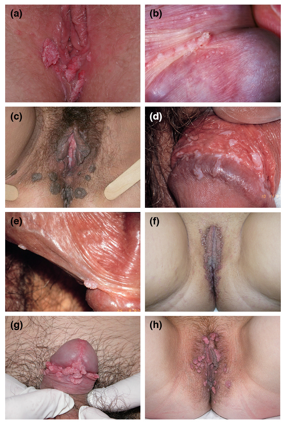
Typical presentations of AGW are shown in Fig. 1. AGW appear as papillomatous plaques or flat lesions and can be single or multiple in number. Lesions vary from flesh‐coloured to white, pink or brown.3 They typically manifest in areas of the body that are in close contact during sex: mainly on the anogenital areas such as vulva, penis, groin, perineum, perianal skin, but also in the oral cavity.2 Diagnosis of clinically typical AGW does not require histological confirmation.
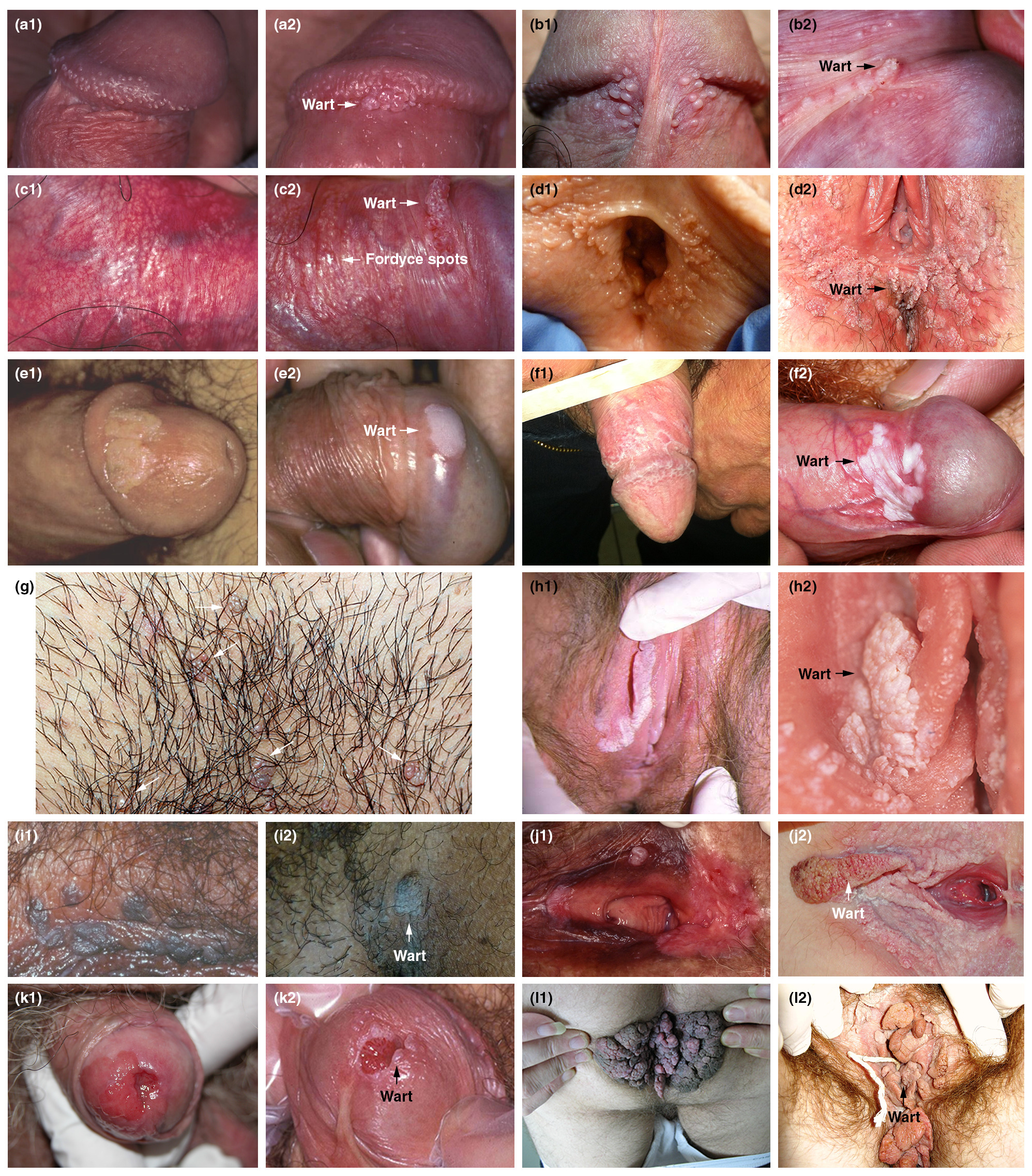
There are many conditions that can be misinterpreted as AGW (Fig. 2). Differential diagnoses that need to be excluded include normal skin variations (e.g. pearly penile papules, parafrenular glands, Fordyce spots, vestibular papillae, sebaceous cysts), other infectious or inflammatory conditions and other papules (syphilis on mucosal plates, molluscum contagiosum, lichen planus, psoriasis, condyloma lata) and benign or malignant neoplastic lesions (papillomatoses of vulva, nevi, verrucous carcinoma, invasive carcinoma, seborrhoeic keratosis, Bowen's disease, Buschke‐Löwenstein disease, pigmented or unpigmented grade 2–3 intraepithelial neoplasia, lymphangioma).2, 3 Pigmented or unusual lesions should be immediately referred to a specialist.
A checklist for the initial consultation with the patient is provided in Table 1. This will help physicians when diagnosing AGW to get the relevant information from the patient in order to manage and treat the AGW effectively. A number of questions that physicians are frequently asked are shown in Table 2, along with suggested answers. Patients should be reassured that if they have developed AGW, appropriate treatment can clear the warts within 3 months.5 Patients should be informed that AGW are of mostly sexual origin and are caused by HPV which is contagious; therefore, it is important for patients to disclose their AGW to recent sexual partners, who should be advised to visit a physician if they have developed AGW. Physicians should also inform patients that smokers have a 27% increased risk of developing AGW compared with non‐smokers.15 Furthermore, they should explain that HPV prevalence in patients who smoke is 48.2% compared with 37.5% for non‐smokers (P < 0.001).16 Generally, warts develop within weeks or months after acquiring HPV but in a significant number of cases, the virus can be dormant for months or years before warts emerge.17
| Checklist |
|---|
|
- AGW, anogenital warts.
| Questions | Answers |
|---|---|
| How did I get AGW? | AGW are caused by HPV.1 Usually, HPV is contracted via sexual interactions: indirect acquisition is rare5 |
| What is the risk of HPV transmission? | The risk of HPV transmission is very high (1.6 sexual interactions are enough to get the infection). The infection is very common and the vast majority of people have the virus during their lifetime |
| Is there a treatment? | Discuss the modalities and the limitations of treatment, explaining this will not eradicate the virus |
| Does smoking increase my risk of developing AGW? | Explain that smokers are at an increased risk of developing AGW and therefore, smoking cessation should be encouraged15 |
| How long will I have AGW for? | AGW can recur several times but with appropriate treatment, most warts should clear within 3 months5 |
| Is this the end of my sex life? | Reassure the patient that this is not the case |
| Should I disclose to my current and previous partner? | It is important to disclose you have AGW to your current partner in order to allow him/her to be checked |
| Should I always use a condom? | Explain that data have shown that increased levels of condom use is associated with increased clearance of HPV.88 It is therefore advisable to use condoms routinely |
| What are the risks during pregnancy? | AGW can become large during pregnancy5 but will usually disappear within weeks of delivery. In rare cases, HPV can be transmitted during child birth resulting in recurrent respiratory papillomatosis in the infant73, 78 |
| Will I develop cancer? | AGW are not related to cancer. AGW are caused by certain types of HPV, other types of HPV can cause cancer5 |
| Can AGW spread to other parts of the body? | It is very uncommon for AGW to spread to other body locations |
- AGW, anogenital warts; HPV, human papillomavirus.
Recommendations when selecting treatment options
Treatment should be individualized for each patient. Although untreated warts can resolve spontaneously,3, 17 most patients want an immediate intervention to eradicate them. Treatments need to be selected on the basis of considerations such as the number, size, morphology, location and keratinization of warts, and whether they are new or recurrent.8, 18 Wart area should be taken into consideration as one study showed that AGW with smaller surface areas (2–19 mm2) require significantly fewer treatment episodes and take less time to clear than those with larger surface areas (100–1038 mm2).19 Patient‐related considerations also need to be taken into account such as their preference for home or clinic‐based treatment, and the convenience of the regimen in terms of dosing frequency and duration.8, 18 Patient‐applied options are often preferred as they offer privacy, convenience and autonomy.18
Treatment options for AGW are provided in Table 3,20-64 and individual modalities are discussed in more detail below. A recent meta‐analysis of 18 studies of patient‐applied therapies concluded that all are more effective than placebo, although treatments cannot be ranked in terms of efficacy due to a lack of head‐to‐head comparisons.65
| Treatment | Mode of action | Schedule | Clearance rate (%) | Recurrence rate (%) | Advantages | Disadvantages | Refs | |||||
|---|---|---|---|---|---|---|---|---|---|---|---|---|
| Ablative techniques | ||||||||||||
| Cryotherapy | Liquid nitrogen freezes and destroys lesions | Applied directly to lesions; repeat for two or three cycles | 46–96 | 18–39 |
|
| 20, 45-49 | |||||
| CO2 and Nd:YAG laser | Laser vaporizes lesions | Under local anaesthesia, protocol depends on type of laser | 23–95 | 2.5–77 |
|
| 20, 48, 50 | |||||
| Electrocautery | High‐frequency electrical currents cause thermal damage to infected tissue | Under local anaesthesia, base of lesion excised; repeat as required | 35–94 | 20–25 |
|
| 18, 20, 49, 51 | |||||
| Surgery | Scissor or scalpel excision | Under local or general anaesthesia; base of lesion excised | 89–93 | 18–65 |
|
| 52-54 | |||||
| Trichloroacetic acid (33–50%) | Acid induces a chemical burn | One to three times per week; repeat as necessary | 70–100 | 18–36 |
|
| 20, 45, 47, 55 | |||||
| Immunotherapies | ||||||||||||
| Imiquimod 5% | Immunomodulator: stimulates interferon and cytokine production | Three nights per week for up to 16 weeks or longer | 35–75 | 6 |
|
| 20-26, 41, 56-62 | |||||
| Imiquimod 3.75% | Immunomodulator: stimulates interferon and cytokine production | Once daily before bedtime for up to 8 weeks | 19–37 | 15–19 |
|
| 20, 27-29 | |||||
| Sinecatechins 10% and 15% | Inflammatory response modulator | Three times daily for up to 16 weeks | 40–81% | 7–12 |
|
| 20, 30-34 | |||||
| Other topical therapy | ||||||||||||
| Podophyllotoxin 0.5% (alcoholic solution) 0.15% (cream) | Antimitotic agent induces tissue necrosis | Twice‐daily to affected areas for 3 consecutive days per week; discontinue for 4 days; repeat for up to 4 weeks | 45–94 | 11–100 |
|
| 20, 31, 35-40, 42-44, 63 | |||||
| Nitric–zinc complex topical solution | Induces a caustic effect on the wart through mummification and protein denaturation/coagulation action | Once or up to four times; repeat at 2‐week intervals if needed | 90–99 | Not evaluated |
|
| 64 | |||||
- AGW, anogenital warts.
Ablative techniques
Ablative techniques are commonly used by physicians to remove warts in daily practice. However, most are awkward and painful for the patient. The major frustration is the high rate of recurrence with these treatments (see below) and the need for repeat therapeutic interventions. Ablative techniques are associated with a risk of bleeding, tissue destruction, slow wound healing and scarring.44, 66
Cryotherapy
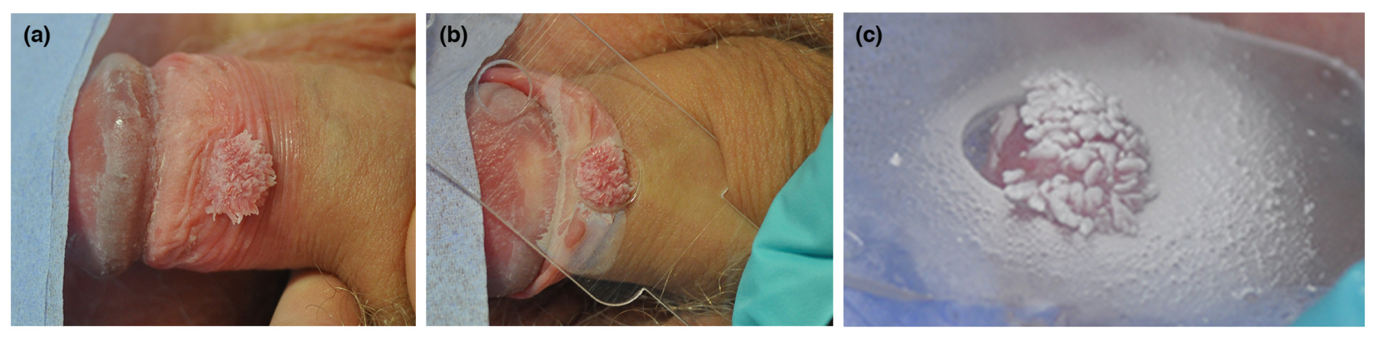
Cryotherapy is the freezing of AGW using liquid nitrogen and is often used at a patient's first clinic visit to help initiate removal of the AGW. Various handheld devices, such as Hydrozid®(Dunelm Pharmaceuticals, Drogheda, Ireland), as well as cryotherapy machines can be used for the procedure. Hydrozid® is a disposable canister, which can be sprayed accurately onto the wart (Fig. 3). This treatment option can be repeated weekly, biweekly or every 3 weeks and is a relatively simple, inexpensive technique, requiring minimal training. However, it requires many clinic visits and a second or third cycle of freezing may be needed. Clearance rates of 46–96% have been reported although treatment can cause pain, necrosis and blistering.45-49, 66 For non‐Caucasians, post‐inflammatory hypo/hyperpigmentation after treatment with cryotherapy can be frustrating; therefore, this should be discussed with patients before proceeding with this treatment option.
Carbon dioxide and Nd:YAG laser
Carbon dioxide (CO2) and Nd:YAG lasers vaporize lesions using focused infrared light energy; however, it is not always possible to know the extent of the infected tissue, and therefore, vaporizing large regions around the warts is not always feasible. Local anaesthesia is usually required, especially on extensive and thick lesions as it can penetrate deeply into the lesions.50
This treatment option is used less frequently than other therapies as it requires specialized and costly equipment, and has an increased risk of serious complications unless used by an experienced physician.1 However, clearance rates of up to 95% have been reported in clinical studies, with a head‐to‐head comparison showing greater efficacy than cryotherapy.47, 48, 50 It is important to note that fumes from laser treatment contain contagious particles and adequate measures should be taken to prevent the virus from spreading. Masks and smoke evacuators should therefore be used.
Electrocautery
Electrocautery uses high‐frequency electrical currents to destroy AGW and requires local anaesthesia and physician expertise.18, 20 Clinical studies have shown clearance rates of 35–94%.20, 49, 51 As fumes from electrocautery contain contagious particles, preventative measures should be put in place to stop the virus spreading.
Surgery
Surgery is performed using scissors or a scalpel and is particularly suited for removing large lesions causing obstruction. Local or general anaesthesia is required, and patients may experience post‐operative pain.50 Clearance rates of up to 93% have been reported in clinical studies.51-53
Trichloroacetic acid (TCA; 33–50%)
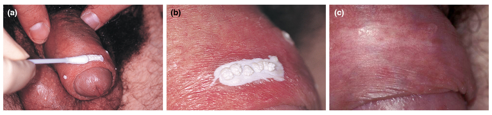
Physician‐applied acidic treatment causes a chemical burn that destroys the AGW. The acid can be administered up to three times per week until the warts have cleared. This process requires a skilled professional to choose the appropriate lesion and duration of application but it is easy to apply and effective for treating AGW, with clearance rates of 70–100% reported in clinical studies (Fig. 4). However, side‐effects such as local discomfort, burning and ulceration are common, hence the need for careful application.45-47, 53, 55, 66 TCA can also be used to treat small lesions; however, it is not frequently used due to a high recurrence rate and the risk of side‐effects.
Immunotherapies
Immunotherapies use stimulation of the body's own immune system to clear infected lesions.
Imiquimod 5% or 3.75%
Imiquimod is an immune response modifier with antiviral activity. This Toll‐like receptor 7 agonist induces the production of cytokines, which enhance the ability of antigen presenting cells to present viral antigens to reactive T lymphocytes.21, 22, 65 Imiquimod 5% has been approved for the treatment of AGW worldwide, whereas imiquimod 3.75% is only approved in certain countries such as the United States of America (USA) and Canada. Imiquimod 5% is self‐applied by the patient three nights per week for up to 16 weeks; if no improvement has occurred after 4–6 weeks, treatment can be applied daily. In comparison, imiquimod 3.75% is self‐applied once‐nightly for up to 8 weeks. Imiquimod 5% may be applied for longer durations if there is a good clinical result but complete clearance has not occurred at the end of the initial treatment period.28 Both imiquimod formulations are associated with local skin reactions such as erythema, pruritus, burning, pain and sometimes erosions. These are all signs that the immune system has been activated. The lower concentration of imiquimod in the 3.75% cream is associated with improved tolerability. In addition, the shorter treatment duration and dosing simplicity may improve patients' adherence to the regimen.27 AGW clearance rates from clinical studies range from 35 to 75% with the 5% cream20, 23, 24, 41, 56-62 and 19 to 37% with the 3.75% formulation,20, 27-29 with higher clearance rates in women than men.29 Further studies have shown that patients find imiquimod 5% to be both acceptable and preferable to other AGW treatments. A study of 559 patients with AGW reported excellent, very good or good with imiquimod 5% in 27.4%, 36.1% and 23.0% of patients, respectively.25 In addition, a survey of 629 patients showed that imiquimod 5% was rated better in terms of overall satisfaction, convenience, time to clearance and lack of associated pain than other AGW therapies.26
Sinecatechins
Sinecatechins consist of green tea polyphenols, which have anti‐inflammatory, anti‐proliferative, pro‐apoptotic and antiviral properties, although their exact mode of action is unknown.30, 31 They are available for the treatment of AGW as a 10% and 15% ointment or cream, which is self‐applied by the patient three times per day for a maximum of 16 weeks.31 In comparison, imiquimod 5% is applied three times weekly while application of imiquimod 3.75% is once daily.30 Patient adherence to dosing regimens should be considered, as compliance is important in achieving treatment effectiveness.30 An additional factor that may affect compliance is that sinecatechin 15% ointment is a brown formulation,67 which could stain light‐coloured clothing and bedding, reducing patient adherence.
Clinical studies of sinecatechins have shown similar clearance rates to that of imiquimod 5% therapy. Sinecatechins have resulted in complete clearance rates of 40–81%, with comparable differences in response rates between the 10% and 15% ointments.31-34Furthermore, the recurrence rate with sinecatechin 10% ointment was 6.8% after 12 weeks of treatment68 and 12% with sinecatechin 10% cream following 12 weeks of treatment.32This was higher than the recurrence rate of 6.2% observed with imiquimod 5% treatment after 3 months and 6.3% at 6 months.62 No significant difference in clearance or recurrence rates has been found between sinecatechin 10% cream and placebo.30 No long‐term data are available for sinecatechins. The most commonly observed application site reactions are erythema, pruritus, irritation, pain and ulceration; these side‐effects may indicate the greater likelihood of a clinical response.30
Other topical therapies
Podophyllotoxin 0.15% cream or 0.5% alcoholic solution
Podophyllotoxin stops division of infected cells causing tissue necrosis.20, 31, 35 It can be self‐applied by patients twice‐daily for three consecutive days, separated by a 4‐day treatment‐free period and repeated for up to 4 weeks. Patients need to carefully apply the solution to the lesions and avoid contact with healthy skin. Clearance rates from clinical studies range from 45 to 94%, with common side‐effects including pain, itching, burning, erosion and inflammation.20, 31, 35-40, 42-44, 63
Nitric zinc
Nitric–zinc complex is a solution for topical application containing nitric acid, zinc, copper and organic acids, currently used to treat common warts.64 It has a caustic effect on the wart through mummification and protein denaturation or a coagulation action.64 The solution can be applied topically once, or up to four times, at 2‐week intervals until a complete clinical cure rate is observed.64 Clearance rates in one study ranged from 90 to 99%, and the product was well tolerated with no serious adverse events recorded.64 Initial data suggest promising efficacy in AGW; however, additional studies are needed.
Guidance for preventing the recurrence of AGW
Anogenital warts recurrence is common and frustrating for patients and physicians.18Recurrence rates with conventional ablative techniques are relatively high (Table 3), since these methods only remove the visible wart without affecting the underlying HPV infection.44, 66 Of currently available treatments, recurrence rates are very low with immunotherapies, imiquimod (6–19%)20, 23, 28, 41, 56, 57, 60, 62 and sinecatechins (4–12%),32-34 as these treatments stimulate the host's immune response to clear the warts.
Studies have shown that a combination of ablative techniques followed by immunotherapy may lead to even lower recurrence rates; ablation provides rapid clearance but has high recurrence rates while immunotherapy has slow clearance rates and a lower risk of recurrence.20, 69 A study of 211 patients showed that imiquimod 5% applied within 3 weeks after laser therapy (to ensure complete wound healing) was associated with a low rate of wart recurrences of 11.8% over 6 months of follow‐up.69 Results of a 3‐arm, open‐label study involving 358 patients showed that 6‐month recurrence rates in those randomized to a combination of ablation followed by imiquimod 5% (8%) were lower than those after ablation alone (26%), but similar to imiquimod 5% monotherapy (6%).62 Furthermore, the results of a retrospective case series of 27 patients showed that combined treatment with cryotherapy, podophyllin 25% and subsequent use of sinecatechins 15% ointment led to a recurrence rate of 7.4% after 6 months of follow‐up.70 Gilson et al.,71 further showed that a combination of cryotherapy and podophyllotoxin cream 0.15% resulted in a higher clearance rate (60%) than with cryotherapy alone (45.7%) at both 4 and 12 weeks. However, these differences were not statistically significant.71
Pre‐treatment of AGW with imiquimod to stimulate an immune reaction followed by surgery is also associated with low recurrence rates. A retrospective study of 60 patients with anogenital warts showed that the recurrence rate during long‐term follow‐up (up to 7 years) was lower for patients with complete responses to imiquimod 5% monotherapy (15%), or with surgical removal of residual warts after imiquimod 5% (20%), compared with surgery alone (65%).54
A simplified algorithm for AGW treatment
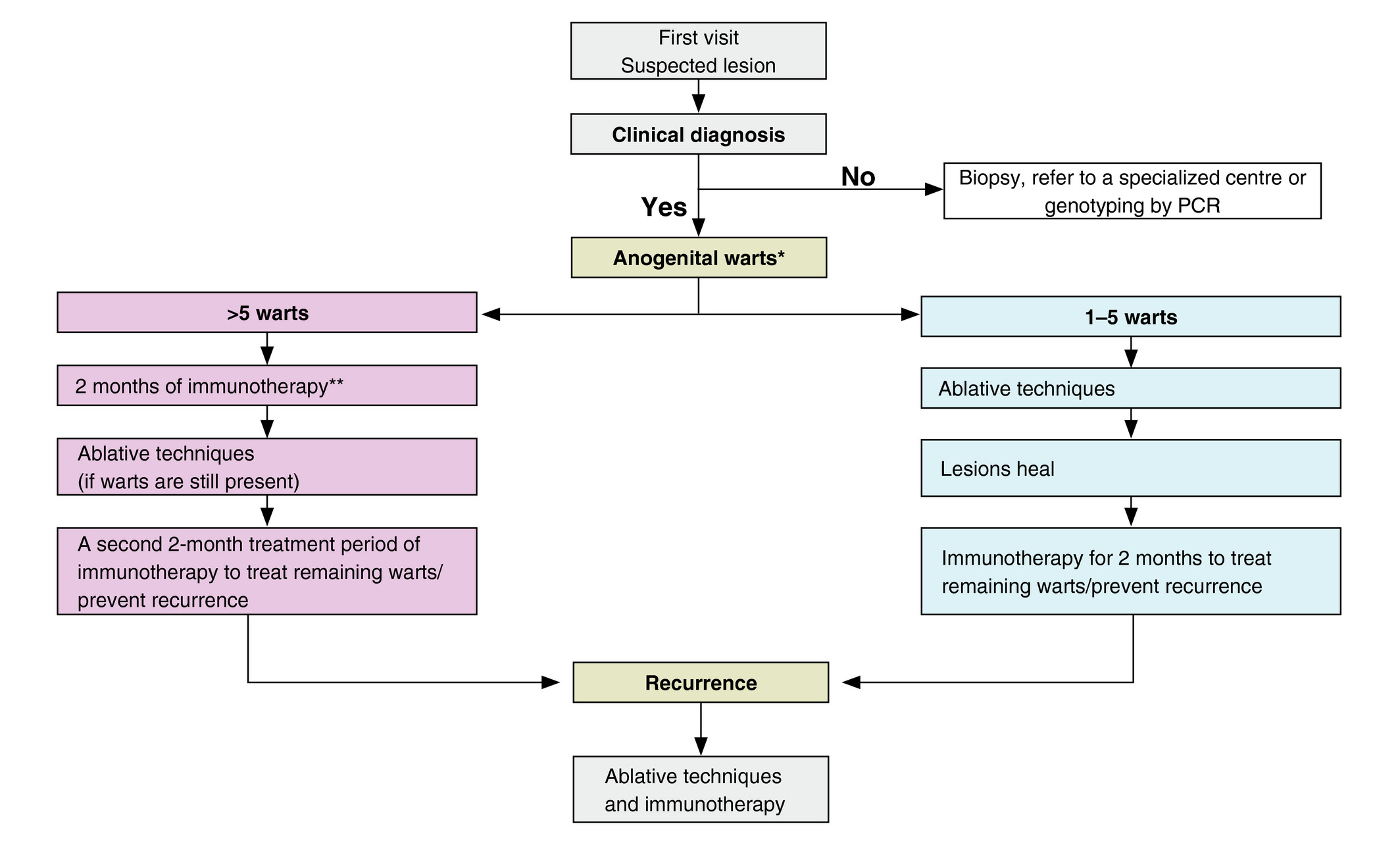
A new simplified treatment algorithm for AGW is shown in Fig. 5. Patients with a confirmed clinical diagnosis of AGW are initially classified by their number of warts. Patients with 1–5 warts may be treated in the first instance with ablation. Once the lesions have healed, immunotherapy can be used for 2 months to treat remaining warts and/or prevent recurrence. The choice of ablative technique is at the discretion of the physician taking factors such as the location of the wart into consideration. For those with more than five warts, the expert's recommendation is to pre‐treat the AGW with an immunotherapy for 2 months to see whether an immune response can be stimulated. If the warts are still present following this treatment, an ablative technique can be used to remove the AGW. It is recommended to use a second 2‐month course of immunotherapy to treat remaining warts and/or prevent recurrence. It is recognized that there are many different algorithms for the treatment of AGW and that the choice is dependent on many factors. For example, if all staff are experienced in ablative techniques, then irrespective of the number of warts, the clinic protocol may dictate that ablation is used on all patients with warts at first visit, followed by immunotherapy. In the UK, this is usual practice as it is preferable for a reduced number of clinic visits.
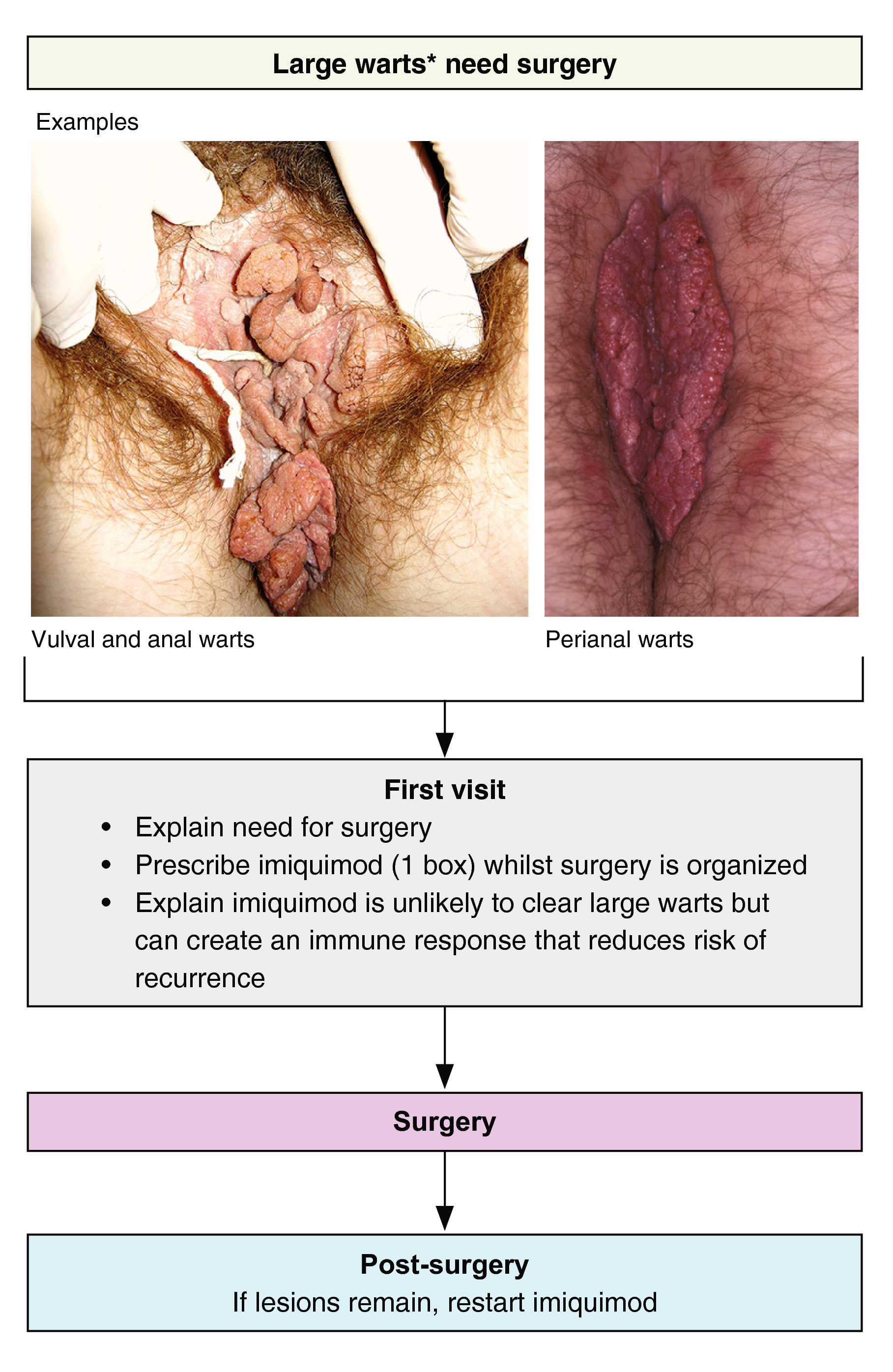
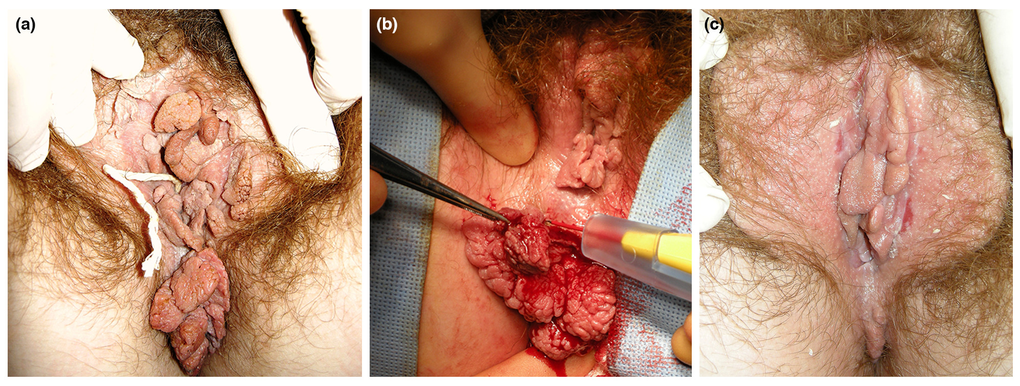
An algorithm for the treatment of patients with large AGW is also shown in Fig. 6. Large warts are defined as too large for local TCA or cryotherapy, and patients with these warts should be referred to a specialist. Based on clinical experience, our recommendation is to initially pre‐treat the AGW with immunotherapy for up to 16 weeks to stimulate an immune reaction to reduce the risk of recurrence. In support of this, long‐term recurrence rates are lower for patients pre‐treated with imiquimod 5% followed by surgery compared with surgery alone.54, 70 Evidence for other immunotherapies in this setting is not currently available. The AGW should then be surgically removed under general anaesthesia, with immunotherapy being re‐started if there are residual or recurrent lesions. A histological examination of the excised tissue should be performed to exclude verrucous or squamous cell carcinoma. An example of a patient treated with this approach is shown in Fig. 7.
Guidance for daily practice situations
Guidance for daily practice situations and the subsequent action that can be taken are shown in Table 4.3, 8, 12, 16, 23, 26, 30, 50, 54, 56, 62, 66, 72-80
| Daily practice situations | Actions |
|---|---|
| AGW remaining following ablation |
|
| Experience or fear of local side‐effects in genital area |
|
| Limited initial efficacy with imiquimod | |
| Lack of adherence |
|
| Lumps left may not be true warts |
|
| Heavy cigarette smoking | |
| Pregnancy |
|
| Immune suppression |
|
| Other conditions (i.e. diabetes, eczema, psoriasis) |
|
| Concomitant local infections (e.g. bacterial, fungal etc.) |
|
Preventing AGW
Anogenital warts can now be effectively prevented using the quadrivalent (HPV 6, 11, 16 and 18) or nanovalent (HPV 6, 11, 16, 18, 31, 33, 45, 52 and 58) HPV vaccines; these protect against HPV types that cause AGW, cervical cancer and other types of anogenital and oral cancer. The HPV quadrivalent vaccine has shown to be up to 100% effective in preventing AGW in association with vaccine‐type HPV in women.81, 82 After its introduction in Australia, a study with a 4‐year follow‐up showed a 59% reduction in the prevalence of AGW in young females.83 There was also a concomitant, although less marked, decline in AGW in heterosexual men following introduction of the vaccine.83, 84 Prevention of AGW with the HPV vaccine could therefore result in substantial savings in healthcare costs and reduction in workload for sexual health clinics.85 The vaccine is also effective in 12‐ to 15‐year‐old boys and is licensed for use in both sexes in most countries where it is available.85, 86 Evidence on whether the vaccination could be useful in AGW treatment is not yet clear; however, there are scientific data supporting use of the vaccination in individuals previously exposed to HPV.87
Conclusions
The guidance provided will help physicians with the diagnosis and management of AGW in daily clinical practice, in order to improve the health and quality of life of patients with AGW. The suggested therapeutic approach is flexible, allowing physicians to choose treatment depending on local availability and physician expertise, as well as considering patient preferences.
Acknowledgements
The authors were assisted in the preparation of the manuscript by David Harrison, Medscript Ltd, and Laura Brennan, a professional medical writer at CircleScience, an Ashfield Company, part of UDG Healthcare plc. Medical writing support was funded by Meda Pharma S.p.A. a Mylan Company. The authors would like to thank Professor Parent (Department of Dermatology, Hôpital Erasme, Université Libre de Bruxelles, Brussels, Belgium) for contributing photographs to this manuscript.
References
- 1 . Warts (genital). BMJ Clin Evid 2010; 2010: 1602.
- 2 , , et al. Clinical features of external genital warts. J Cutan Med Surg 2013; 17( Suppl 2): S55– S60.
- 3 . New approach to managing genital warts. Can Fam Physician 2013; 59: 731– 736.
- 4 , , et al. Understanding genital warts: epidemiology, pathogenesis, and burden of disease of human papillomavirus. J Cutan Med Surg 2013; 17( Suppl 2): S47– S54.
- 5 , . Frequently asked questions about genital warts in the genitourinary medicine clinic: an update and review of recent literature. Sex Transm Infect 2008; 84: 3– 7.
- 6 . Genital warts: current and future management options. Am J Clin Dermatol 2005;6: 239– 243.
- 7 , , et al. Psychological burden of anogenital warts. J Eur Acad Dermatol Venereol 2009; 23: 1035– 1038.
- 8 , , et al. Managing external genital warts: practical aspects of treatment and prevention. J Cutan Med Surg 2013; 17( Suppl 2): S68– S75.
- 9 , . Follow‐up of men with genital papilloma virus infection. Psychosexual aspects. Acta Derm Venereol 1992; 72: 185– 186.
- 10 , , et al. Economic and humanistic burden of external genital warts. Pharmacoeconomics 2012; 30: 1– 16.
- 11 , , et al. The costs of managing genital warts in the UK by devolved nation: England, Scotland, Wales and Northern Ireland. Int J STD AIDS 2016; 27: 51– 57.
- 12 , , et al. 2012 European guideline for the management of anogenital warts. J Eur Acad Dermatol Venereol 2013; 27: e263– e270.
- 13 , , et al. UK national guidelines on the management of anogenital warts 2015. URL http://www.bashh.org/documents/UK%20national%20guideline%20on%20Warts%202015%20FINALpdf. (last accessed: 26 February 2018).
- 14 Centers for Disease Control and Prevention. Sexually transmitted diseases treatment guidelines: Anogenital warts. 2015 URL http://www.cdc.gov/std/tg2015/warts.htm. (last accessed: 26 February 2018)
- 15 , , et al. Association between smoking and genital warts: longitudinal analysis. Sex Transm Infect 2010; 86: 258– 262.
- 16 , , . The impact of smoking on HPV infection and the development of anogenital warts. Int J Colorectal Dis 2014; 29: 899– 908.
- 17 , , . Genital warts: a comprehensive review. J Clin Aesthet Dermatol 2012; 5: 25– 36.
- 18 , , et al. Therapeutic options for external genital warts. J Cutan Med Surg 2013; 17( Suppl 2): S61– S67.
- 19 , , . Factors involved in clearance of genital warts. Int J STD AIDS2001; 12: 789– 792.
- 20 , , et al. Sinecatechins and imiquimod as proactive sequential therapy of external genital and perianal warts in adults. Int J STD AIDS 2017; 28: 1433– 1443.
- 21 . Immunomodulatory and pharmacologic properties of imiquimod. J Am Acad Dermatol 2000; 43: S6– S11.
- 22 . Mechanism of action and other potential roles of an immune response modifier. Cutis 2007; 79: 36– 45.
- 23 , , et al. Imiquimod, a patient‐applied immune‐response modifier for treatment of external genital warts. Antimicrob Agents Chemother 1998; 42: 789– 794.
- 24 , , et al. Efficacy and safety of imiquimod versus podophyllotoxin in the treatment of anogenital warts. Sex Transm Dis 2011; 38: 216– 218.
- 25 , . Effectiveness, satisfaction and compliance with imiquimod in the treatment of external anogenital warts. Int J STD AIDS 2003; 14: 11– 17.
- 26 , , et al. New patient‐applied therapy for anogenital warts is rated favourably by patients. Int J STD AIDS 2001; 12: 565– 570.
- 27 , . The role of imiquimod 3.75% cream in the treatment of external genital warts. Skin Therapy Lett 2012; 17: 5– 7.
- 28 , , et al. Imiquimod 3.75% cream applied daily to treat anogenital warts: combined results from women in two randomized, placebo‐controlled studies. Infect Dis Obstet Gynecol 2011; 2011: 806105.
- 29 , , . Imiquimod cream 2.5% and 3.75% applied once daily to treat external genital warts in men. Cutis 2015; 96: 277– 282.
- 30 , . Sinecatechins 10% ointment: a green tea extract for the treatment of external genital warts. Skin Therapy Lett 2015; 20: 6– 8.
- 31 , . Sinecatechins (Polyphenon E) ointment for treatment of external genital warts and possible future indications. Expert Opin Biol Ther 2014; 14: 1033– 1043.
- 32 , , et al. A randomized, double‐blind, four‐arm parallel‐group, placebo‐controlled Phase II/III study to investigate the clinical efficacy of two galenic formulations of Polyphenon E in the treatment of external genital warts. J Eur Acad Dermatol Venereol 2007; 21:1404– 1412.
- 33 , , et al. Topical Polyphenon E in the treatment of external genital and perianal warts: a randomized controlled trial. Br J Dermatol 2008; 158: 1329– 1338.
- 34 , , et al. Sinecatechins, a defined green tea extract, in the treatment of external anogenital warts: a randomized controlled trial. Obstet Gynecol 2008; 111:1371– 1379.
- 35 , . Management of external genital warts. Am Fam Physician 2014; 90:312– 318.
- 36 , , et al. A single‐blind study of podophyllotoxin cream 0.5% and podophyllotoxin solution 0.5% in male patients with genital warts. Genitourin Med 1995; 71:391– 392.
- 37 , , et al. Self‐treatment of female external genital warts with 0.5% podophyllotoxin cream (Condyline) vs weekly applications of 20% podophyllin solution. Int J STD AIDS 1995; 6: 257– 261.
- 38 , , et al. An open, comparative, study of the efficacy of 0.5% podophyllotoxin lotion and 25% podophyllotoxin solution in the treatment of condylomata acuminata in males and females. Int J STD AIDS 1993; 4: 194– 199.
- 39 , . Self‐treatment using a 0.5% podophyllotoxin cream of external genital condylomata acuminata in women. A placebo‐controlled, double‐blind study. Sex Transm Dis 1992;19: 170– 174.
- 40 , , et al. A double‐blind, randomized trial of 0.5% podofilox and placebo for the treatment of genital warts in women. Obstet Gynecol 1991; 77: 735– 739.
- 41 , , et al. Self‐administered topical 5% imiquimod cream for external anogenital warts. HPV Study Group. Human PapillomaVirus. Arch Dermatol 1998; 134: 25– 30.
- 42 , , et al. Self‐treatment using 0.25–0.50% podophyllotoxin‐ethanol solutions against penile condylomata acuminata: a placebo‐controlled comparative study. Genitourin Med 1994; 70: 105– 109.
- 43 , , et al. Patient‐applied podofilox for treatment of genital warts. Lancet 1989; 1: 831– 834.
- 44 , , et al. Double‐blind randomized clinical trial of self‐administered podofilox solution versus vehicle in the treatment of genital warts. Am J Med 1990; 88: 465– 469.
- 45 , , et al. Cryotherapy compared with trichloroacetic acid in treating genital warts. Genitourin Med 1987; 63: 390– 392.
- 46 , . Cryotherapy in the treatment of condylomata acuminata: a controlled study of 64 patients. J Dermatol Surg Oncol 1991; 17: 273– 276.
- 47 , , . Treatment of external genital warts comparing cryotherapy (liquid nitrogen) and trichloroacetic acid. Sex Transm Dis 1993; 20: 344– 345.
- 48 , , . CO(2) Laser therapy versus cryotherapy in treatment of genital warts; a Randomized Controlled Trial (RCT). Iran J Microbiol 2012; 4: 187– 190.
- 49 , , et al. Treatment of external genital warts: a randomised clinical trial comparing podophyllin, cryotherapy, and electrodesiccation. Genitourin Med 1990; 66:16– 19.
- 50 , . An evidence‐based review of medical and surgical treatments of genital warts. Dermatol Online J 2006; 12: 5.
- 51 , , et al. Randomized clinical trial comparing systemic interferon with diathermocoagulation in primary multiple and widespread anogenital condyloma. Obstet Gynecol 1989; 74: 393– 397.
- 52 . Comparison of podophyllin application with simple surgical excision in clearance and recurrence of perianal condylomata acuminata. Lancet 1985; 2: 1146– 1148.
- 53 . Podophyllin versus scissor excision in the treatment of perianal condylomata acuminata: a prospective study. Br J Surg 1989; 76: 1067– 1068.
- 54 , , . Treatment of anogenital warts with imiquimod 5% cream followed by surgical excision of residual lesions. J Am Acad Dermatol 2002; 47: S212– S216.
- 55 , , et al. Therapeutic value of trichloroacetic acid in the treatment of isolated genital warts on the external female genitalia. J Reprod Med 2007; 52: 521– 525.
- 56 , , et al. Treatment of genital warts with an immune‐response modifier (imiquimod). J Am Acad Dermatol 1998; 38: 230– 239.
- 57 , , et al. Imiquimod 5% cream is a safe and effective self‐applied treatment for anogenital warts–results of an open‐label, multicentre Phase IIIB trial. Int J STD AIDS 2001; 12: 722– 729.
- 58 , , et al. Safety and efficacy of imiquimod 5% cream in the treatment of penile genital warts in uncircumcised men when applied three times weekly or once per day. Int J STD AIDS 2001; 12: 22– 28.
- 59 , , et al. Treatment of external genital warts in men using 5% imiquimod cream applied three times a week, once daily, twice daily, or three times a day. Sex Transm Dis 2001; 28: 226– 231.
- 60 , , et al. Topical imiquimod 5% cream in external anogenital warts: a randomized, double‐blind, placebo‐controlled study. J Dermatol 2004; 31: 627– 631.
- 61 , , et al. An open‐label phase II pilot study investigating the optimal duration of imiquimod 5% cream for the treatment of external genital warts in women. Int J STD AIDS 2006; 17: 448– 452.
- 62 , , et al. Randomized, comparative trial on the sustained efficacy of topical imiquimod 5% cream versus conventional ablative methods in external anogenital warts. Eur J Dermatol 2006; 16: 642– 648.
- 63 , , . Podophyllotoxin 0.5% v podophyllin 20% to treat penile warts. Genitourin Med 1988; 64: 263– 265.
- 64 , , et al. Efficacy and tolerability of nitric‐zinc complex in the treatment of external genital warts and "difficult‐to‐treat" warts: a "proof of concept", prospective, multicentre, open study. G Ital Dermatol Venereol 2015; 150: 643– 648.
- 65 , , et al. Self‐administered interventions for anogenital warts in immunocompetent patients: a systematic review and meta‐analysis. Sex Transm Infect2017; 93: 155– 161.
- 66 . Evaluation of imiquimod for the therapy of external genital and anal warts in comparison with destructive therapies. Br J Dermatol 2007; 157( Suppl 2): 52– 55.
- 67 . Update on the treatment of genital warts. Dermatol Online J 2013; 19: 18559.
- 68 , , et al. Polyphenon E: a new treatment for external anogenital warts. Br J Dermatol 2010; 162: 176– 184.
- 69 , , et al. Effect of adjuvant imiquimod 5% cream on sustained clearance of anogenital warts following laser treatment. Infect Dis Obstet Gynecol 2002;10: 79– 88.
- 70 , , et al. Combined treatment of anogenital HPV infection with cryodestruction, podophyllin 25% and post‐ablation immunomodulation with sinecatechins 15% ointment – a retrospective analysis. Int J STD AIDS 2016; 27: 1071– 1078.
- 71 , , et al. A multicentre, randomised, double‐blind, placebo controlled study of cryotherapy versus cryotherapy and podophyllotoxin cream as treatment for external anogenital warts. Sex Transm Infect 2009; 85: 514– 519.
- 72 , , et al. 5% Imiquimod cream for external anogenital warts in HIV‐infected patients under HAART therapy. Int J STD AIDS 2004; 15: 17– 20.
- 73 , , et al. Recurrent respiratory papillomatosis: A state‐of‐the‐art review. Respir Med 2017; 126: 116– 121.
- 74 , , et al. Effects of nicotine on the immune response. II. Chronic nicotine treatment induces T cell anergy. J Immunol 1996; 156: 2384– 2390.
- 75 , , . Young women's experiences of managing self‐treatment for anogenital warts. Sex Reprod Healthc 2012; 3: 117– 121.
- 76 , , . Skin infections in pregnancy. Clin Dermatol 2016; 34: 368– 377.
- 77 , , . Imiquimod use in the genital area and development of lichen sclerosus and lichen planus. Int J STD AIDS 2010; 21: 219– 221.
- 78 , , . Recurrent respiratory papillomatosis. Otolaryngol Clin North Am 2012; 45: 671– 694, viii‐ix.
- 79 , . Topical imiquimod: a review of its use in the management of anogenital warts, actinic keratoses, basal cell carcinoma and other skin lesions. Drugs 2007; 67:2187– 2210.
- 80 , , et al. Diabetes and genital warts: an unhappy coalition. Int J STD AIDS 2010; 21: 457– 459.
- 81 , , et al. Four year efficacy of prophylactic human papillomavirus quadrivalent vaccine against low grade cervical, vulvar, and vaginal intraepithelial neoplasia and anogenital warts: randomised controlled trial. BMJ 2010; 341: c3493.
- 82 , , et al. Quadrivalent vaccine against human papillomavirus to prevent anogenital diseases. N Engl J Med 2007; 356: 1928– 1943.
- 83 , , et al. Quadrivalent human papillomavirus vaccination and trends in genital warts in Australia: analysis of national sentinel surveillance data. Lancet Infect Dis2011; 11: 39– 44.
- 84 , , et al. Rapid decline in presentations of genital warts after the implementation of a national quadrivalent human papillomavirus vaccination programme for young women. Sex Transm Infect 2009; 85: 499– 502.
- 85 , . HPV vaccination–reaping the rewards of the appliance of science. BMJ2013; 346: f2184.
- 86 , , et al. Efficacy of quadrivalent HPV vaccine against HPV Infection and disease in males. N Engl J Med 2011; 364: 401– 411.
- 87 , , et al. Recurring infection with ecologically distinct HPV types can explain high prevalence and diversity. Proc Natl Acad Sci U S A 2017; 114: 13573– 13578.
- 88 , , et al. Association of condom use, sexual behaviors, and sexually transmitted infections with the duration of genital human papillomavirus infection among adolescent women. Arch Pediatr Adolesc Med 2006; 160: 151– 156.
-
Chem. Commun. , 2019, Accepted Manuscript DOI : 10.1039/C8CC10001B, Communication Ning Dong, Hao Luo, Yonggao Xia, Haocheng Guo, Rushe...
-
Inorganic Chemistry DOI: 10.1021/acs.inorgchem.8b03235 from Journals via hj on Inoreader http://bit.ly/2CAsmuf
-
Painful right elbow mass Perspective from a pediatric orthopedic deformity surgeon Alkaptonuria—an atypical case: multi-modality imaging rev...

