Monday, November 14, 2022

TMIC-78. CIRCADIAN REGULATOR CLOCK DRIVES IMMUNOSUPPRESSION IN GLIOBLASTOMA

alexandrossfakianakis shared this article with you from Inoreader
Abstract
The symbiotic interactions between cancer stem cells and the tumor microenvironment (TME) are critical for tumor progression. However, the molecular mechanism underlying this symbiosis in glioblastoma (GBM) remains enigmatic. Here, we show that circadian locomotor output cycles kaput (CLOCK) and its heterodimeric partner brain and muscle ARNT-like 1 (BMAL1) in glioma stem cells (GSCs) drive immunosuppression in GBM. Integrated analyses of the data from transcriptome profiling, single-cell RNA sequencing, and TCGA datasets, coupled with functional studies, identified legumain (LGMN) as a direct transcriptional target of the CLOCK–BMAL1 complex in GSCs. Moreover, CLOCK-directed olfactomedin-like 3 (OLFML3) upregulates LGMN in GSCs via the hypoxia-inducible factor 1-alpha (HIF1A) signaling. Consequently, LGMN promotes microglial infiltration into the GBM TME via upregulating CD162, and polarizes infiltrating microglia towards an immune-suppressive phenotype. In GBM mouse models, inhibition of the CLOCK–OLFML3–HIF1A–LGMN–CD162 axis reduces intratumoral immune-suppressive microglia, increases CD8+ T cell infiltration, activation and cytotoxicity, and synergizes with anti-PD1 therapy. In human GBM, the CLOCK-regulated LGMN signaling correlates positively with microglial level and poor prognosis. Together, these findings uncover the CLOCK–OLFML3–HIF1A–LGMN axis as a molecular switch that controls microglial biology and immunosuppression, thus revealing potential new therapeutic targets for GBM patients.
View on Web

CSIG-19. THE PI3K/AKT/MTOR SIGNALING CASCADE MAY CONTRIBUTE TO SEX DIFFERENCES IN GLIOBLASTOMA

alexandrossfakianakis shared this article with you from Inoreader
Abstract
Glioblastoma (GBM) is the most common and aggressive primary brain tumor in adults. It is more prevalent in males and female patients exhibit a survival advantage. Understanding the molecular mechanisms that underlie those sex differences could support novel treatment strategies. The PI3K/Akt/mTOR signaling cascade plays a crucial role in GBM development and progression as it is involved in the regulation of cellular growth, survival, nutrient sensing, and metabolic activity. To investigate whether the PI3K/Akt/mTOR signaling cascade may differ in male and female GBM patients, we first assessed survival data of male and female GBM patients using the TCGA RPPA phosphoproteome data set and found that changes in protein phosphorylation of components of the PI3K/Akt/mTOR pathway, including mTOR phosphorylation, worsen the outcome of male but not female GBM patients in a dose-dependent fashion. This was supported by in vitro experiments of murine tran sformed astrocytes where pathway stimulation via treatment with insulin, IGF-1, or EGF significantly increased pathway activity in male but not female cells. Furthermore, pathway inhibition via serum deprivation resulted in a significant decrease in pathway activity in male but not female cells, indicating that male transformed astrocytes exhibit higher sensitivity to inhibitory conditions (serum deprivation) and stimulatory conditions (insulin, IGF-1, or EGF treatment). Together, these data suggest that (i) the PI3K/Akt/mTOR pathway activity may affect male and female GBM outcome differently, and (ii) there are sex differences in the regulation of the PI3K/Akt/mTOR signaling cascade in GBM which may contribute to the sex disparity in GBM. Our data add to the growing body of literature regarding the presence of sex differences in PI3K/Akt/mTOR signaling in health and disease and provide important insight for the development of translatable approaches to treatment for male and female GBM patients.
View on Web

Varicella Zoster Virus Reactivation Following COVID‐19 Vaccination in patients with autoimmune inflammatory rheumatic diseases: A cross‐sectional Chinese study of 318 cases

alexandrossfakianakis shared this article with you from Inoreader

Abstract

Recently, varicella-zoster virus (VZV) reactivation has been observed after the administration of coronavirus disease 2019 (COVID-19) vaccines. Autoimmune inflammatory rheumatic diseases (AIIRDs) patients are at a higher risk for VZV reactivation for immunocompromised status. The study aimed to investigate the adverse events (AEs), especially VZV reactivation, following vaccination against SARS-CoV-2 in a Chinese cohort of AIIRD patients. A cross-sectional survey using an online questionnaire was conducted among AIIRD patients and healthy controls (HCs). Multivariate logistic regression was used to identify potential factors associated with VZV reactivation. 318 AIIRD patients and 318 age and sex-matched HCs who got COVID-19 inactivated vaccines were recruited. The main AIIRDs are rheumatoid arthritis (31.8%) and systemic lupus erythematous (23.9%). Most of patients (85.5%) had stable disease and 13.2% of them had aggravation after vaccination. Compared to H Cs, patients had higher rates of rash (p=0.001), arthralgia (p<0.001) and insomnia (p=0.007). In addition, there were 6 (1.9%) AIIRD patients and 5 (1.6%) HCs reported VZV reactivation after the COVID-19 vaccination (p=0.761). Multivariate logistic regression analysis illustrated that diabetes mellitus (OR, 20.69; 95%CI, 1.08-396.79; p=0.044), chronic hepatitis B virus infection (OR, 24.34; 95%CI, 1.27-466.74; p=0.034), and mycophenolate mofetil (OR, 40.61; 95%CI, 3.33-496.15; p=0.004) independently identified patients with VZV reactivation. Our findings showed that the inactivated COVID-19 vaccination was safe for AIIRD patients though some patients could suffer from VZV reactivation.

This article is protected by copyright. All rights reserved.

View on Web

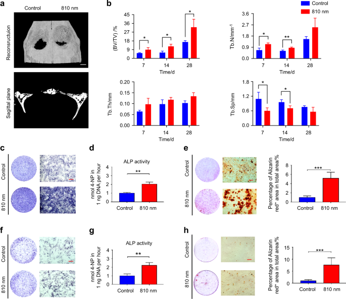
Low intensity near-infrared light promotes bone regeneration via circadian clock protein cryptochrome 1

alexandrossfakianakis shared this article with you from Inoreader

41368_2022_207_Fig1_HTML.png

International Journal of Oral Science, Published online: 14 November 2022; doi:10.1038/s41368-022-00207-y

Low intensity near-infrared light promotes bone regeneration via circadian clock protein cryptochrome 1
View on Web

Sunday, November 13, 2022

Comparison of passive versus active transcutaneous bone anchored hearing devices in the pediatric population

alexandrossfakianakis shared this article with you from Inoreader
Transcutaneous bone anchored hearing devices (BAHDs) were introduced in an effort to avoid potential complications associated with the abutment of percutaneous BAHDs. Transcutaneous BAHDs can be active or pass...
View on Web

Exhaled Mycobacterium tuberculosis Predicts Incident Infection in Household Contacts

alexandrossfakianakis shared this article with you from Inoreader

m_ciac455f1.jpeg?Expires=1731185268&Sign

Abstract
BackgroundHalting transmission of Mycobacterium tuberculosis (Mtb) by identifying infectious individuals early is key to eradicating tuberculosis (TB). Here we evaluate face mask sampling as a tool for stratifying the infection risk of individuals with pulmonary TB (PTB) to their household contacts.
Methods
Forty-six sputum-positive PTB patients in The Gambia (August 2016–November 2017) consented to mask sampling prior to commencing treatment. Incident Mtb infection was defined in 181 of their 217 household contacts as QuantiFERON conversion or an increase in interferon-γ of ≥1 IU/mL, 6 months after index diagnosis. Multilevel mixed-effects logistical regression analysis with cluster adjustment by household was used to identify predictors of incident infection.
Results
Mtb was detected in 91% of PTB mask samp les with high variation in IS6110 copies (5.3 × 10² to 1.2 × 10⁷). A high mask Mtb level (≥20 000 IS6110 copies) was observed in 45% of cases and was independently associated with increased likelihood of incident Mtb infection in contacts (adjusted odds ratio, 3.20 [95% confidence interval, 1.26–8.12]; P = .01), compared with cases having low-positive/negative mask Mtb levels. Mask Mtb level was a better predictor of incident Mtb infection than sputum bacillary load, chest radiographic characteristics, or sleeping proximity.
Conclusions
Mask sampling offers a sensitive and noninvasive tool to support the stratification of individuals who are most infectious in high-TB-burden settings. Our approach can provide b etter insight into community transmission in complex environments.
View on Web

Ultrasonographic Features of Salivary Glands after Radioiodine Therapy in Patients with Thyroid Cancer

alexandrossfakianakis shared this article with you from Inoreader
Ultrasonographic Features of Salivary Glands after Radioiodine Therapy in Patients with Thyroid Cancer

Chronic sialadenitis is the most common radioactive iodine (RAI) treatment complication. We explored the sonographic features of the major salivary glands in patients with papillary thyroid cancer (PTC) treated with RAI. RAI treatment following thyroidectomy is associated with a dose-response effect and adverse changes in the major salivary glands and should be prescribed carefully.


Objective

Chronic sialadenitis is the most common complication of radioactive iodine (RAI) treatment. The aim of the study was to ascertain sonographic features of the major salivary glands in patients with papillary thyroid cancer (PTC) treated with RAI.

Methods

The database of a tertiary medical center was retrospectively searched for consecutive patients who underwent total thyroidectomy for PTC in 2011–2020 with ultrasound follow-up after 1 year. Changes in ultrasound features of the major salivary glands were compared between patients treated or not treated with RAI postoperatively.

Results

The cohort included 158 patients, of whom 109 (69%) were treated postoperatively with RAI (mean dose, 131 mCi) and 49 were not (control group). Sonographic changes were observed in the major salivary glands in 43% of the study group and 18% of the control group (p = 0.002), including coarse echotexture, decreased echogenicity, fibrosis, and atrophy. Higher RAI doses were significantly correlated with the prevalence and severity of glandular changes (p < 0.0001).

Conclusion

RAI treatment following thyroidectomy is associated with a dose–response effect and adverse changes in the major salivary glands and should be prescribed carefully.

Level of Evidence

4 Laryngoscope, 2022

View on Web

Sensory neuromuscular electrical stimulation for dysphagia rehabilitation: A literature review

alexandrossfakianakis shared this article with you from Inoreader

Abstract

Background

Dysphagia is a common disorder following a cerebrovascular accident. It can cause detrimental effects on patient's quality of life and nutrition intake, especially in older adults. Neuromuscular electrical stimulation has been one of the management strategies for acceleration of the recovery. This review summarizes the current evidence on sensory threshold stimulation of the procedure.

Method

This review compiled data from the Internet database PubMed, Cochrane Library and Scopus using combination of MeSH thesaurus: "Sensory threshold", "electrical stimulation", "neuromuscular stimulation", "Deglutition", "Dysphagia". 11 studies were intergraded into the review.

Results

Most of the studies show significant improvement to the outcomes of sensory neuromuscular electrical stimulation treatment. In many cases, the results of the treatment are comparable or superior to motor threshold stimulation and conventional therapy. However, the study design and parameters of the procedure varied greatly without conclusive standardized guidelines.

Conclusion

The sensory neuromuscular electrical stimulation (SNMES) is a viable treatment option for treating oropharyngeal dysphagia. The most suggested application parameters are an intensity at sensory threshold, a frequency of 80 Hz, an impulse time of 700 μs, a combined total duration of 20 hours of stimulation in a 2-week period, placing the electrodes in the submental area of the neck. However, further research is necessary to construct a definitive guideline for clinicians.

View on Web

Evaluating the role of non-alcoholic fatty liver disease in cardiovascular diseases and type 2 diabetes: a Mendelian randomization study in Europeans and East Asians

alexandrossfakianakis shared this article with you from Inoreader

m_dyac212f1.jpeg?Expires=1731265321&Sign

Abstract
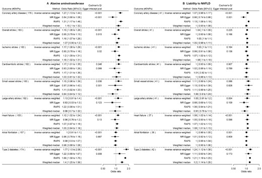
BackgroundWhether non-alcoholic fatty liver disease (NAFLD) causes cardiovascular disease (CVD) and type 2 diabetes (T2D) is unclear and possible differences between ethnicities have not been thoroughly explored. We used Mendelian randomization (MR) to assess the role of NAFLD in CVD and T2D risk in Europeans and East Asians.
Methods
We conducted a MR study using genetic predictors of alanine aminotransferase (ALT), liability to NAFLD, aspartate transaminase (AST), liver magnetic resonance imaging corrected T1 and proton density fat fraction and combined them with genome-wide association studies (GWAS) summary statistics of CVD, T2D and glycaemic traits (sample size ranging from 14 400 to 977 320). Inverse-variance weighted analysis was used to assess the effect of NAFLD in these outcomes, with sensitivity analyses and replication in FinnGen. We conducted analyses in East Asians using ethnicity-specific genetic predictors of ALT and AST, and the respective outcome GWAS summary statistics.
Results
In Europeans, higher ALT was associated with higher T2D risk (odds ratio: 1.77 per standard deviation, 95% CI 1.5 to 2.08), with similar results for other exposures, across sensitivity analyses and in FinnGen. Although NAFLD proxies were related to higher coronary artery disease (CAD) and stroke risk, sensitivity analyses suggested possible bias by horizontal pleiotropy. In East Asians, higher ALT was possibly associated with higher T2D risk, and ALT and AST were inversely associated with CAD.
Conclusions
NAFLD likely increases the risk of T2D in Europeans and East Asians. Potential differential effects on CAD between Europeans and East Asians require further investigation.
View on Web

Neutrophil‐to‐lymphocyte and hypopharyngeal cancer prognosis: System review and meta‐analysis

alexandrossfakianakis shared this article with you from Inoreader

Abstract

Several studies have reported the value of neutrophil-to-lymphocyte ratio (NLR) for the prognosis of hypopharyngeal cancer. However, contradictory findings have also been published. We aimed to clarify the effect of NLR on the prognosis of hypopharyngeal cancer through meta-analysis. Systematic search of PubMed and other database with study selection and data extraction. The combined hazard ratio (HR) and 95% confidence intervals (CI) were calculated using STATA, applying either a fixed-effects or random-effects model. Meta-regression, subgroup analysis, and sensitivity analysis were used to analyze sources of heterogeneity. Publication bias were also assessed. This meta-analysis included 2232 patients with hypopharyngeal cancer from seven studies. The combined HR (OS, HR = 1.80, 95CI%, 1.14–2.82; PFS, HR = 1.88, 95CI%, 1.26–2.79) suggested that high NLR was associated with poor overall survival (OS) and progression-free survival (PFS). Pretreatment NLR can be used as an effective serological indicator to assess the prognosis of patients with hypopharyngeal cancer.

View on Web

Collaboration request

Hi there How would you like to earn a 35% commission for each sale for life by selling SEO services Every website owner requires the ...