Monday, May 16, 2022

Temporomandibular disorder symptoms in young adults: Three‐dimensional impact on oral health‐related quality of life

alexandrossfakianakis shared this article with you from Inoreader

Abstract

Objectives

This study examined the three-dimensional impact of pain-related and/or intra-articular Temporomandibular disorder (TMD) symptoms and ascertained the inter-relationships between the functional, pain, and psychosocial aspects of oral health-related quality of life (OHRQoL).

Methods

Young adults were enlisted from a local university and TMD symptoms were assessed with the Diagnostic Criteria for TMDs Symptom Questionnaire (DC/TMD-SQ). Participants were stratified into those with no (NT), pain-related (PT), intra-articular (IT), and mixed (MT) TMD symptoms. OHRQoL was examined using the Oral Health Impact Profile-14 (OHIP-14), and dimensional effects (oral function [OF], orofacial pain [OP], and psychosocial impact [PI]) were established subsequently. Data were evaluated with Kruskal-Wallis, Dunn, and Wilcoxon signed-rank tests (α = 0.05).

Results

The mean age of the participants (n = 1205) was 19.7 ± 1.3 years (71.8% women). While 42.2% reported no TMD symptoms, 22.3%, 16.9%, and 18.5% had PT, IT, and MT accordingly. The MT, PT, and IT groups presented higher total OHIP-14 and dimension scores than the NT group (p <0.01). Scores were also significantly different between the MT and IT groups except for OF. For all TMD groups, the ranking of dimensional impact was OP > PI > OF, and associations between the OP and PI dimensions were the strongest (correlation coefficient [r s ] = 0.57-0.76).

Conclusions

Young adults with PT and/or IT symptoms have poorer OHRQoL, especially in the OP and PI dimensions, which were moderate to strongly correlated. The use of the three dental Patient-reported Outcome (dPRO) dimensions could provide construct equivalency among OHRQoL measures.

View on Web

Ergonomics in Otolaryngology: A Systematic Review and Meta‐analysis

alexandrossfakianakis shared this article with you from Inoreader
Ergonomics in Otolaryngology: A Systematic Review and Meta-analysis

Ergonomic stressors among otolaryngologists contribute to a high rate of WRMD across all subspecialties with notable impact on productivity, longevity, and quality of life. The overall proportion (95% confidence intervals [CI]) of general otolaryngologists reporting WRMD was 0.79 (0.66, 0.88) for any symptoms; 0.54 (0.40, 0.67) for neck symptoms; 0.33 (0.20, 0.49) for shoulder symptoms; and 0.49 (0.40, 0.59) for back symptoms.


Objectives

To determine the proportion of otolaryngologists with work-related musculoskeletal discomfort (WRMD) and to review objective ergonomic data that contribute to WRMD.

Study Design

Systematic review and meta-analysis.

Methods

A comprehensive search of the literature identified 1121 articles for initial review of which 19 (3563 participants) met criteria for qualitative discussion and eight (2192 participants) met criteria for meta-analysis. Random effects meta-analyses were used to estimate the proportion of otolaryngologists reporting WRMD.

Results

The overall proportion (95% confidence intervals [CI]) of general otolaryngologists reporting WRMD was 0.79 (0.66, 0.88) for any symptoms; 0.54 (0.40, 0.67) for neck symptoms; 0.33 (0.20, 0.49) for shoulder symptoms; and 0.49 (0.40, 0.59) for back symptoms. Surgeons performing primarily subspecialty cases had a lower estimated overall prevalence of WRMD versus those performing general ENT cases, however the odds ratio (OR) was not statistically significant (OR [95% confidence interval] 0.53 [0.22, 1.25]). 23%–84% of otolaryngologists underwent medical treatment for WRMD. 5%–23% took time off work and 1%–6% stopped operating completely as a result of WRMD. 23%–62.5% of otolaryngologists believed WRMD negatively impacted their quality of life. Objective measures of ergonomic posture indicate moderate to severe risk of injury during the routine clinic and surgical procedures with none found to be low risk.

Conclusions

Ergonomic stressors among otolaryngologists contribute to a high rate of WRMD across all subspecialties with notable impact on productivity, longevity, and quality of life. Laryngoscope, 2022

View on Web

Pembrolizumab und Cetuximab als therapeutische Optionen für adenoidzystische Karzinome des Kopf-Hals-Bereiches

alexandrossfakianakis shared this article with you from Inoreader

lro-1054_10-1055-a-1830-8298-1.jpg

Laryngorhinootologie
DOI: 10.1055/a-1830-8298


Georg Thieme Verlag KG Rüdigerstraße 14, 70469 Stuttgart, Germany

Article in Thieme eJournals:
Table of contents  |  Full text

View on Web

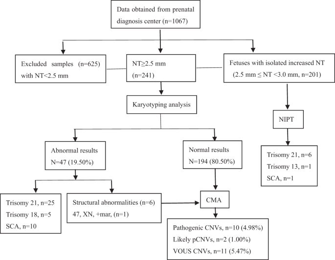
Chromosomal microarray analysis versus noninvasive prenatal testing in fetuses with increased nuchal translucency

alexandrossfakianakis shared this article with you from Inoreader

10038_2022_1041_Fig1_HTML.png

Journal of Human Genetics, Published online: 17 May 2022; doi:10.1038/s10038-022-01041-0

Chromosomal microarray analysis versus noninvasive prenatal testing in fetuses with increased nuchal translucency
View on Web

Radiographic bone volume alteration after jaw cyst enucleation with or without simultaneous bone grafts: A prospective randomized study

alexandrossfakianakis shared this article with you from Inoreader

Abstract

Objective

This study was aimed to evaluate bone healing after jaw cyst enucleation with or without bone substitutes by cone beam computed tomography, and to analyze potential influence factors for bone formation as well.

Materials and Methods

Sixty seven jaw cyst patients were randomly assigned to two groups. Thirty three patients in control group accepted cystectomy without any filling material. The rest 34 bone cavities which filled with xenograft (DBBM, Bio-Oss®) and covered by absorbable membrane (Bio-Gide®) were included in the guided bone regeneration (GBR) group. All patients were examined with cone bean computerized tomography before operation, 3 and 6 months after surgery. Linear regression analysis was applied to evaluate the influence factors of bone healing.

Results

There was no significant difference in bone formation rate at 3 months after enucleation, with shrinkage rate (SR) of cystic lesion in control group and GBR group of 26.43 ± 14.98% and 20.78 ± 10.80%, respectively (p > 0.05). Larger shrinkage area in GBR group was detected on postoperative radiographs after 6 months with SR of 60.11 ± 19.23%, when compared to those in patients without filling (6 months SR: 48.63 ± 19.39%, p = 0.018, <0.05). Linear regression analysis showed that cyst size was negatively correlated with bone formation.

Conclusion

GBR with bovine xenograft and absorbable membrane showed considerable bone regeneration property in the healing of jaw cystic defects after enucleation of radicular cysts. Cyst size showed a suppressive influence on bone formation.

View on Web

Sunday, May 15, 2022

Type 2 laryngeal cleft associated with OpitzG/BBB syndrome

alexandrossfakianakis shared this article with you from Inoreader

1-s2.0-S2173573522X00045-cov150h.gif

Publication date: May–June 2022

Source: Acta Otorrinolaringologica (English Edition), Volume 73, Issue 3

Author(s): Cristina García-Muro, Isabel Sáenz-Moreno, Fernando Gómez-Sáez, Ana Navazo-Eguia

View on Web

Survival Rate of Ultrawide Diameter Implants Placed Into Molar Postextraction Sockets and in Function for Up to 144 Months

alexandrossfakianakis shared this article with you from Inoreader

Abstract

Purpose

Innovations in macroimplant design, specifically ultrawide implants 7.0 mm or greater in diameter, have allowed immediate molar replacement. This is a retrospective study assessing the survival rates of ultrawide diameter implants (7.0, 8.0, 9.0 mm) immediately placed into molar extraction sockets. Implants were followed up to 144 months post-placement.

Materials and methods

A retrospective study was conducted of all patients treated in a private surgical practice between January 1, 2008, and December 31, 2020, who received ultrawide dental implants (7.0, 8,0, 9.0mm.) placed immediately into molar extraction sockets. Wide diameter healing abutments were placed on all implants at the time of surgery. Abutments and crown restorations were fabricated after at least 4 months of unloaded healing. Patient age, gender, implant location and implant diameters were examined for survival. Insertion torque values at the time of placement and time in function were also evaluated. Biometric statistics were computed with P values (<0.05. Descriptive and bivariate statistics were computed; P values were set at .05.

Results

: Five hundred forty-four patients (225 males; 319 females) [average age 62.5 years; range 27 to 95] had 563 implants placed. Five hundred thirty-five of five hundred sixty-three (535/563) implants survived; 28 failed [clinical survival rate (CSR) 95.03%]. Number and time in function were: 0–12 years 100%; 0–9 years 85%; 0–6 years 69%; 0–3 years 35% or 10–12 years 16%; 7–9 years 16%; 4–6 years 34%; 0–3 years 35%. No significant differences were found between gender and implant failures (p = 0.22). Maxillary (266/285; 93.3%) and mandibular (269/278; 96.8%) implant CSRs were not significantly different. Three implant diameters were used: 7.0 mm (206/563) [36.6%]; 8.0 mm (267/563) [47.4%]; 9.0 mm (90/563) [15.9%]. Clinical survival rates were: 7.0 mm (201/206) [97.6%]; 8.0 mm (252/267) [94.4%]; 9.0 mm (82/90) [91.1%]. Mean age for patients with failed implants did not show any significant differences (p = 0.1398). Fifteen of the 28 failed implants failed within 120 da ys of surgical placement (prior to definitive restoration; [53.6%]; 4 implants failed between 4 and 12 months [14.3%]; 9 implants failed at least 1-year post-loading [32.1%].

Conclusions

The results of this long-term retrospective study regarding ultrawide diameter implants suggested that these implants were viable treatment options for immediate molar replacement following tooth extraction in either jaw with an unloaded healing protocol. High clinical survival rates were reported over a 144 month (12 year) timeframe.

This article is protected by copyright. All rights reserved

View on Web

Mucosal Melanoma of the Oral Cavity: What is the Role of Elective Neck Dissection?

alexandrossfakianakis shared this article with you from Inoreader
Mucosal Melanoma of the Oral Cavity: What is the Role of Elective Neck Dissection?

This study uses the National Cancer Database to analyze factors associated with survival in oral cavity mucosal melanoma, as well as the role of elective neck dissections. Lymph node involvement, distant metastasis, age, race, and male sex were associated with worse outcomes. While elective neck dissections did not improve overall survival, they may have a prognostic role and help select patients for treatment intensification.


Objectives

Mucosal melanoma (MM) is a rare malignancy that can present in the head and neck (H&N). The Oral cavity is the second most common primary site in the H&N after sinonasal mucosa. This study investigates the impact of demographic and clinical factors on survival in oral cavity MM. Further, it investigates the outcomes and utility of elective neck dissections (END) in the management of oral MM.

Methods

The National Cancer Database was used to evaluate 432 patients with oral cavity MM from 2004 to 2016. Kaplan-Meir and Cox regression analyses were used to determine variables associated with survival.

Results

The mean age was 64.0 ± 16.0 years. Most patients were white (85.1%) and male (60.0%). Gingiva (37.6%) and hard palate (36.1%) were the most common primary subsites in the oral cavity. Five-year overall survival was 31.0%. Age (Hazards Ratio [95% Confidence Interval], 1.03 [1.01–1.06]), N-stage (1.94 [1.10–3.42]), M-stage (10.13 [3.33–30.86]), male sex (1.79 [1.06–3.03]), and African-American race (2.63 [1.14–6.11]) were significantly associated with worse survival. 199 patients (46.9%) underwent neck dissection including 118 with lymph node yield (LNY) ≥ 18. The rate of occult nodal positivity was 45.4% for LNY ≥ 18 and 28.3% for LNY ≥ 1. ENDs were not associated with improved outcomes. However, occult lymph node involvement was associated with worse overall survival (p = 0.004).

Conclusions

Oral cavity MM has a poor prognosis. Lymph node involvement, distant metastasis, age, race, and male sex are associated with worse outcomes. Performing an END did not improve survival. However, END may have a prognostic role and help select patients for treatment intensification.

Level of Evidence

4 Laryngoscope, 2022

View on Web

Systematic Review of CT Angiography in Guiding Management in Pediatric Oropharyngeal Trauma

alexandrossfakianakis shared this article with you from Inoreader
Systematic Review of CT Angiography in Guiding Management in Pediatric Oropharyngeal Trauma

The utility of CT angiography (CTA) in children with minor appearing oropharyngeal trauma is controversial. This article is a systematic review of the changes in diagnosis and treatment based on CTA results in pediatric patients with oropharyngeal trauma.


Objectives

Pediatric oropharyngeal trauma is common. Although most cases resolve uneventfully, there have been reports of internal carotid artery injury leading to devastating neurovascular sequelae. There is significant controversy regarding the utility of CT angiography (CTA) in children with seemingly minor oropharyngeal trauma. The goal of this study was to appraise changes in diagnosis and treatment based on CTA results.

Methods

A comprehensive search of PubMed, Embase, CINAHL, Scopus, the Cochrane Ear, Nose and Throat Disorders Group Trials Register, and the ClinicalTrials.gov database was performed following PRISMA guidelines.

Results

The search yielded 5,078 unique abstracts, of which 8 articles were included. A total of 662 patients were included, with 293 having any CT head/neck imaging, and 255 with CTA. Eleven injuries/abnormalities of the carotid were found on CTAs, comprising edema around the carotid (n = 8), potential intimal tear (n = 1), carotid spasm (n = 1), and carotid compression (n = 1). The pooled proportion of imaging findings on CTA that could lead to changes in clinical management was 0.00 (95% CI 0.00–0.43). Angiography was obtained in 10 patients, in 6 cases due to abnormal CTA. Angiography identified 1 patient with vessel spasm and two patients with carotid intima disruption without thrombus. No patient underwent vascular repair or suffered cerebrovascular injury.

Conclusion

Imaging with CTA yielded radiological abnormalities in a few instances. These results do not support the routine use of CTA in screening pediatric oropharyngeal trauma when balanced against the risk of radiation, as it rarely resulted in management changes and was not shown to improve outcomes.

Level of Evidence

N/A Laryngoscope, 2022

View on Web

Comparison of Adult and Pediatric Cochlear Implant Wound Complications: A Meta‐Analysis

alexandrossfakianakis shared this article with you from Inoreader
Comparison of Adult and Pediatric Cochlear Implant Wound Complications: A Meta-Analysis

The objective of this study is to compare age-related differences in wound complications following cochlear implantation (CI). CI wound complication rates reported in the literature are low; however, adults have a higher risk of these complications than pediatric patients. The reported complication rate in elderly adults is low.


Objective

To compare age-related differences in wound complications following cochlear implantation (CI).

Methods

We performed a systematic review of PubMed, Cochrane Database, and Web of Science databases to identify original research evaluating the patient-level factors (demographics and medical history) associated with wound complications following CI. Outcomes were expressed as relative risk (RR) with 95% confidence intervals using the inverse variance method. Studies without comparison groups were described qualitatively.

Results

Thirty-eight studies representing 21,838 cochlear implantations were included. The rate of wound complications ranges from 0% to 22%. Patient age (adult versus pediatric) was the only factor with comparison groups appropriate for meta-analysis. The 10 studies (n = 9547 CI's) included in the meta-analysis demonstrated that adults had a higher incidence of overall wound complications (2.94%) than in children (2.44%) (RR 1.31, 95% CI 1.01–1.69). Adults had a higher incidence of general/unclassified wound complications (2.07%) than in children (1.34%) (RR 1.68, 95% CI 1.12–2.52). There was no difference between adults and children for specific complications such as hematoma, infection, or seroma. Elderly patients (over age 75) have wound complication rates that range from 1% to 4%. No studies contained comparison groups regarding other patient-level factors and CI wound complications.

Conclusion

CI wound complication rates reported in the literature are low; however, adults have a higher risk of these complications than pediatric patients. The reported complication rate in elderly adults is low. There is a gap in CI research in consistently reporting wound complications and rigorous research investigating the impact of patient-level factors and wound complications.

Level of Evidence

Not applicable Laryngoscope, 2022

View on Web

Collaboration request

Hi there How would you like to earn a 35% commission for each sale for life by selling SEO services Every website owner requires the ...