Thursday, January 14, 2021

Soft Palate Fistula in a Patient With Normal Palate as a Rare Complication After Septoplasty

xloma.fota13 shared this article with you from Inoreader

SocialThumb.00001665.DC.jpeg

While the formation of a palatal fistula after septoplasty has been reported previously, it is a rare occurrence, especially in a patient with a normal palate. In most of the previous reports, the palatal fistulas were located on the hard palate and associated with various underlying conditions. Here, we present a case of soft palate fistula which developed in a patient with a normal palate after septoplasty. The 20-year old woman complained of liquid regurgitation after her septoplasty procedure. A pin-point size fistula opening was observed on the soft palate. A shallow depression was identified at the junction between the hard and soft palate by a computed tomography scan done before surgery. The fistula was subsequently repaired through a palatal flap. After surgery, the defect was completely closed, and the patient had no more symptoms. In addition, we review the previously reported cases of palatal fistula after septoplasty. Address correspondence and reprint requests to Ji-Hyeon Shin, MD, PhD, Department of Otolaryngology-Head and Neck Surgery, College of Medicine, The Catholic University of Korea, 222 Banpo-daero, Seocho-gu, Seoul, 06591, Republic of Korea; E-mail: tachyon0217@gmail.com Received 1 September, 2020 Accepted 21 November, 2020 The author reports no conflicts of interest. © 2021 by Mutaz B. Habal, MD.
View on the web

Augmentation Genioplasty Using a Third Molar as a Bone Graft: An Alternative Surgical Technique

xloma.fota13 shared this article with you from Inoreader

SocialThumb.00001665.DC.jpeg

Purpose: Augmentation genioplasty corrects vertical chin deficiency. To stabilize the synthesis and to allow ossification, a biomaterial prop is necessary. Third molars are frequently removed during orthognathic surgery and provides good material used as autogenous grafts. Surgical Technique: This article describes the surgical technique of an augmentation genioplasty using a third molar as a prop biomaterial. Results are stable in time. This technique presents no risk of infection and is less invasive than other autogenous bone graft. Conclusions: third molar can be used as a prop biomaterial in augmentation genioplasty Address correspondence and reprint requests to Dr Adeline Kerbrat, Hopital Pitié Salpétrière, Service de Stomatologie et Chirurgie Maxillo-faciale, 81 Boulevard de l'hopital, 75013 Paris, France; E-mail: adelinekerbrat@hotmail.com Received 28 August, 2020 Accepted 4 December, 2020 The authors report no conflicts of interest. © 2021 by Mutaz B. Habal, MD.
View on the web

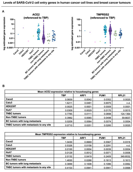
Cancers, Vol. 13, Pages 296: Triple-Negative Breast Cancer and the COVID-19 Pandemic: Clinical Management Perspectives and Potential Consequences of Infection

xloma.fota13 shared this article with you from Inoreader
Via Cancers

cancers-13-00296-g001-550.jpg

Cancers, Vol. 13, Pages 296: Triple-Negative Breast Cancer and the COVID-19 Pandemic: Clinical Management Perspectives and Potential Consequences of Infection

Cancers doi: 10.3390/cancers13020296

Authors: Justin M. Brown Marie-Claire D. Wasson Paola Marcato

The COVID-19 pandemic has caused the need for prioritization strategies for breast cancer treatment, where patients with aggressive disease, such as triple-negative breast cancer (TNBC) are a high priority for clinical intervention. In this review, we summarize how COVID-19 has thus far impacted the management of TNBC and highlighted where more information is needed to hone shifting guidelines. Due to the immunocompromised state of most TNBC patients receiving treatment, TNBC management during the pandemic presents challenges beyond the constraints of overburdened healthcare systems. We conducted a literature search of treatment recommendations for both primary and targeted TNBC therapeutic strategies during the COVID-19 outbreak and noted changes to treatment timing and drugs of choice. Further, given that SARS-CoV-2 is a respiratory virus, which has systemic consequences, management of TNBC patients with metastatic versus localized disease has additional considerations during t he COVID-19 pandemic. Published dataset gene expression analysis of critical SARS-CoV-2 cell entry proteins in TNBCs suggests that the virus could in theory infect metastasized TNBC cells it contacts. This may have unforeseen consequences in terms of both the dynamics of the resulting acute viral infection and the progression of the chronic metastatic disease. Undoubtedly, the results thus far suggest that more research is required to attain a full understanding of the direct and indirect clinical impacts of COVID-19 on TNBC patients.

View on the web

Versatile Approach to Septonasal Deformity: Skyscraper Construction Based on an Old Relic

xloma.fota13 shared this article with you from Inoreader

SocialThumb.00001665.DC.jpeg

Introduction: Severely deformed noses usually harbor a combination of both bony pyramid and septal deformities. In this retrospective study, the authors aimed to evaluate our results of repair in patients with severe nasal deformities and importance of a versatile approach in these cases. Materials and Methods: A total of 32 cases with congenital or acquired (traumas or surgeries) severe nasal deformity were included in this retrospective study. Gender, age, etiology, reconstruction methods, complications, and results were recorded. Preoperative and postoperative pictures were compared; additionally, patients' reviews on the esthetic and functional outcomes were noted. Open approach, weak L-strut template preparation attached to a strong keystone skeleton and reconstruction with a stable L- or T-strut on this template were carried out in all cases. In addition, glabellar flaps were used in 2 cases to restore the contracted skin envelope and wide-angle L-shape cartilage grafts in 7 cases for extensive alar cartilage reconstruction. Results: Favorable esthetic and functional results were obtained in most of the patients. The postoperative problems were recorded as intranasal synechiae; costochondral graft displacement; residual external deviation; nostril asymmetry; residual alar, columellar and tip problems; and prolonged edema. Conclusions: Sufficient sizes and amounts of skin, mucosa, cartilage, and bone tissue must be available to plan versatile repair using flaps and grafts according to the needs of each patient. Preserved stability of the keylock area is substantial. The authors advocate construction of a new structure based on the native weakened skeleton free from the extrinsic and intrinsic forces is an effective method. EBM LEVEL 4 Address correspondence and reprint requests to Omer R. Ozerdem, MD, A-Plast Esthetic Plastic Surgery Center, Akasya Kent Kule A-1 K:24 D:124 34660 Acibadem, Istanbul, Turkey; E-mail: ozerdemor@yahoo.com Received 6 August, 2020 Accepted 4 December, 2020 The authors report no conflicts of interest. © 2021 by Mutaz B. Habal, MD.
View on the web

Self-Cross-Linked Hyaluronic Acid Hydrogel in Endonasal Endoscopic Dacryocystorhinostomy: A Randomized, Controlled Trial

xloma.fota13 shared this article with you from Inoreader

SocialThumb.00001665.DC.jpeg

Purpose: To investigate whether self-cross-linked HA hydrogel fill stimulates wound mucosal regeneration and its epithelialization around the ostia to improve long-term ostial patency in endonasal endoscopic dacryocystorhinostomy (En-DCR). Methods: One hundred and ninety-two patients with unilateral primary chronic dacryocystitis (PCD) were randomized divided into 2 groups: group A (the HA hydrogel group) and group B (the control group). All patients underwent En-DCR. The HA hydrogel group received HA hydrogel filling the ostium at the end of the surgery and the control group received no treatment. The mucosal epithelialization of the wound, the formation of granulation, the formation of scars, and the success rate of ostial patency were compared. Results: Our study included 82 patients in group A and 79 patients in group B. At the 2-week follow up, 74 patients (90.2%) in the group A had a healed ostium with a lining of intact epithelial mucosa. It was higher when compared with 56 patients (70.9%) in group B (X2 = 9.698, P 
View on the web

Lacrimal Duct Obstruction Caused by Nasomaxillary Fracture: A Retrospective Analysis of Consecutive 12 Patients by Computed Tomographic Dacryocystography

xloma.fota13 shared this article with you from Inoreader

SocialThumb.00001665.DC.jpeg

Nasomaxillary fracture is a characteristic mid-facial fracture, and there are no reports showing the fracture damaging the nasolacrimal system (NLS). This report described nasomaxillary fracture cases with NLS damages, which were assessed by computed tomographic dacryocystography (CT-DCG). A retrospective co hort study of nasomaxillary fractures diagnosed by CT was conducted from 2007 to 2015. Twelve patients (mean age: 27.5 years) were found, and their clinical symptoms were as follows: nasal deformity in 10 patients, infra-orbital hypoesthesia in 7, epiphora in 5, and diplopia in one. CT-DCG was performed for 2 patients who complained epiphora, and obstruction was found in 1 patient. All patients underwent open reduction and internal fixation (ORIF), and epiphora in 5 patients was improved. One patient, however, complained epiphora postoperatively, which was supposed to be due to the unsuitable screw insertion. Since nasomaxillary fracture could give NLS obstruction, CT-DCG is useful for diagnosis. Although ORIF is an optimal treatment, attention is needed to avoid the lacrimal canal in screwing on the nasomaxillary buttress. Address correspondence and reprint requests to Yuki Hasegawa, MD, Department of Plastic and Reconstructive Surgery, Tokyo Women's Medical University, 8-1 Kawada-cho, Shinjuku-ku, Tokyo, 162-8666, Japan; E-mail: hasegawa.yuuki@twmu.ac.jp Received 30 June, 2020 Accepted 10 December, 2020 The authors report no conflicts of interest. Supplemental digital contents are available for this article. Direct URL citations appear in the printed text and are provided in the HTML and PDF versions of this article on the journal's Web site (www.jcraniofacialsurgery.com). © 2021 by Mutaz B. Habal, MD.
View on the web

A Posterior Rotational Flap Technique Using Distraction Osteogenesis for Unilateral Lambdoid Craniosynostosis

xloma.fota13 shared this article with you from Inoreader

SocialThumb.00001665.DC.jpeg

Introduction: The craniofacial asymmetry seen in unilateral lambdoid craniosynostosis may not be effectively treated by posterior cranial vault remodeling, endoscopic suturectomy, and helmet therapy, or suturectomy and distraction osteogenesis alone due to limitations in soft-tissue envelope expansion and relapse of the deformity. The authors report a series of unilateral lambdoid craniosynostosis patients treated with a posterior rotational cranial-flap technique using internal distraction osteogenesis. Methods: Posterior cranial vault reconstruction combined with internal distraction was used, aided by preoperative virtual surgical planning. An in situ posterior rotational flap osteotomy was utilized to maximize dural preservation. Primary outcome measures included age-adjusted volume change and age-adjusted percent volume change per mm distraction. Distraction characteristics and perioperative characteristics were also assessed. Results: A total of 5 patients were identified. Mean predistraction intracranial volume was 1087.5 cc (SD  = 202.3 cc) and mean postdistraction included intracranial volume was 1266.1cc (SD  = 131.8cc). Mean age-adjusted percent included intracranial volume change was 14.1% (SD  = 9.6%), and mean percent intracranial volume change per mm distraction was 0.43%/mm distraction (SD  = 0.37%/mm distraction). One patient developed a distractor site infection postoperatively that was treated successfully with oral antibiotics. All patients had a Whitaker score of 1 at one year follow up. Conclusions: Posterior cranial vault remodeling using osteogenesis and a rotational cranial flap technique with dural preservation can be effectively used to maximize bone flap viability and limit postoperative relapse in patients with unilateral lambdoid craniosynostosis. Long term analysis as well as comparison to open techniques will need to be interrogated. Address correspondence and reprint requests to Amanda A. Gosman, MD, UC San Diego/Rady Children's Hospital, Division of Plastic Surgery, 7920 Frost Street, San Diego, CA 92123; E-mail: agosman@health.ucsd.edu Received 24 June, 2020 Accepted 3 December, 2020 The authors report no conflicts of interest. Supplemental digital contents are available for this article. Direct URL citations appear in the printed text and are provided in the HTML and PDF versions of this article on the journal's Web site (www.jcraniofacialsurgery.com). © 2021 by Mutaz B. Habal, MD.
View on the web

Predictors of the Size and Surgical Freedom of the Trans-Cribriform and Trans-Clival Corridors, a Radiographic Analysis

xloma.fota13 shared this article with you from Inoreader

10-1055-s-0040-1722663_200223-1.jpg

J Neurol Surg B Skull Base
DOI: 10.1055/s-0040-1722663

Introduction The transcribriform and transclival corridors are endoscopic endonasal approaches used to treat pathologies of the skull base. We present a predictive model that uses the clival length and ethmoidal width to predict the size and surgical freedom (SF) of these corridors. Methods Adult facial computed tomography scans were reviewed. Exclusion criteria included patients <18 years of age or radiographic evidence of trauma, neoplasm, or congenital deformities of the skull base. The images were analyzed using OsiriX MD (Bernex, Switzerland). Patients' demographics, clival length, ethmoidal width, surface area, and others were collected. Linear regression was used to create prediction models for the size and SF of the transclival and transcribriform corridors. Results A total of 103 patients were included with an average age of 44.9 years and 47% males. Females had a smaller clival surface area (8 vs. 9.2 cm2, p = 0.001). For transclival corridor, clival length correlated positively with SF in the sagittal plane (rho = 0.44, p < 0.05) and negatively with SF in the coronal plane (rho =  − 0.2, p < 0.05). For transcribriform corridor, ethmoidal width correlated positively with SF in the coronal plane (rho = 0.74, p < 0.05), and negatively with SF in the sagittal plane (rho =  − 0.2, p < 0.05). Conclusion A significant variability of the bony anatomy of the anterior and central skull base was found. The use of clival length and ethmoidal width as part of preoperative surgical planning might help to overcome the anatomical variability which could affect the adequacy of surgical corridors.
[...]

Georg Thieme Verlag KG Rüdigerstraße 14, 70469 Stuttgart, Germany

Article in Thieme eJournals:
Table of contents  |  Abstract  |  Full text

View on the web

Anatomical Variations of the Jugular Foramen Region in Patients with Pulsatile Tinnitus

xloma.fota13 shared this article with you from Inoreader

10-1055-s-0040-1722670_200261-1.jpg

J Neurol Surg B Skull Base
DOI: 10.1055/s-0040-1722670

Objective Structural anomalies of the jugular foramen (JF) and adjacent structures may contribute to development of pulsatile tinnitus (PT). The goal of this study was to assess anatomical variants in the ipsilateral JF region in patients with PT and to explore possible predisposing factors for PT. Materials and Methods One hundred ninety-five patients with PT who underwent CT angiography and venography of the temporal bone were retrospectively analyzed. Anatomic variants including dominance of the ipsilateral JF, bony deficiency of the sigmoid sinus and internal carotid artery canal, high riding or dehiscent jugular bulb, dehiscence of the superior semicircular canal, tumors in the JF region, or cerebellopontine angle were assessed. Results Of 195 patients with PT, the prevalence of a dominant JF on the ipsilateral side of patients with PT was 67.2%. Furthermore, the dominant JF demonstrated a significant correlation with the presence of ipsilateral PT (p < 0.001). No anatomical variants were present in 22 patients (11.3%), whereas in patients with structural variants, bony deficiency of the sigmoid sinus was most common (65.6%), followed by high riding (54.9%) or dehiscent jugular bulb (14.4%). Dehiscent internal carotid artery canal (3.1%) and superior semicircular canal (4.1%) were occasionally identified, while arteriovenous fistula, arterial aneurysm and tumors arising from the JF region or cerebellopontine angle were rarely encountered. Conclusion Structural abnormalities of the JF and adjacent structures may predispose to the development of PT. Knowledge of these anatomical variants in the JF region may help establish a clinical strategy for addressing PT.
[...]

Georg Thieme Verlag KG Rüdigerstraße 14, 70469 Stuttgart, Germany

Article in Thieme eJournals:
Table of contents  |  Abstract  |  Full text

View on the web

Aggressive Treatment Including Endonasal Surgical Sequestrectomy with Vascularized Nasoseptal Flap Can Improve Outcomes of Skull Base Osteoradionecrosis

xloma.fota13 shared this article with you from Inoreader

10-1055-s-0040-1722669_200189-1.jpg

J Neurol Surg B Skull Base
DOI: 10.1055/s-0040-1722669

Objective Skull base osteoradionecrosis (SB-ORN) is a serious, potentially lethal complication of radiation therapy. We aimed to review the clinical characteristics and outcomes of SB-ORN according to the extent of treatment. Design Retrospective analysis design was used for this study. Setting The study was conducted in two tertiary care hospitals. Participants Patients included who had been clinically diagnosed with SB-ORN from January 2006 to 2017. Main Outcome Measures Clinical characteristics, including demographics, predisposing factors, presenting symptoms, radiological findings, treatment modalities, and treatment outcomes, were reviewed. Treatment was classified into conservative and aggressive types. Aggressive treatment included radical surgical removal of soft tissue and bony sequestrum with the placement of vascularized tissue. Treatment outcome was analyzed in terms of clinical control, survival, and carotid artery blow out. Results Fifteen patients (11 males and 4 females) were identified during the study period. Eight patients were managed conservatively, whereas seven patients were managed with aggressive treatment. The 2-year survival was 75% in the aggressive treatment group and 15% in the conservative group (log-rank, p = 0.049). The estimated 2-year blow out free rate was 46.7% for the conservative group and 100% for the aggressive group (log-rank, p = 0.100). Conclusion In patients with SB-ORN, aggressive management, including surgical removal of sequestrum and coverage with a pedicled flap, is associated with increased survival.
[...]

Georg Thieme Verlag KG Rüdigerstraße 14, 70469 Stuttgart, Germany

Article in Thieme eJournals:
Table of contents  |  Abstract  |  Full text

View on the web

Cognitive Pathways to Belief in Karma and Belief in God

xloma.fota13 shared this article with you from Inoreader

Abstract

Supernatural beliefs are ubiquitous around the world, and mounting evidence indicates that these beliefs partly rely on intuitive, cross‐culturally recurrent cognitive processes. Specifically, past research has focused on humans' intuitive tendency to perceive minds as part of the cognitive foundations of belief in a personified God—an agentic, morally concerned supernatural entity. However, much less is known about belief in karma—another culturally widespread but ostensibly non‐agentic supernatural entity reflecting ethical causation across reincarnations. In two studies and four high‐powered samples, including mostly Christian Canadians and mostly Hindu Indians (Study 1, N = 2,006) and mostly Christian Americans and Singaporean Buddhists (Study 2, N = 1,752), we provide the first systematic empirical investigation of the cognitive intuitions underlying various forms of belief in karma. We used path analyses to (a) replicate tests of t he previously documented cognitive predictors of belief in God, (b) test whether this same network of variables predicts belief in karma, and (c) examine the relative contributions of cognitive and cultural variables to both sets of beliefs. We found that cognitive tendencies toward intuitive thinking, mentalizing, dualism, and teleological thinking predicted a variety of beliefs about karma—including morally laden, non‐agentic, and agentic conceptualizations—above and beyond the variability explained by cultural learning about karma across cultures. These results provide further evidence for an independent role for both culture and cognition in supporting diverse types of supernatural beliefs in distinct cultural contexts.

View on the web

Collaboration request

Hi there How would you like to earn a 35% commission for each sale for life by selling SEO services Every website owner requires the ...